File:FIG 15.jpg
From EyeWiki
Size of this preview: 800 × 296 pixels. Other resolution: 1,871 × 692 pixels.
Original file (1,871 × 692 pixels, file size: 123 KB, MIME type: image/jpeg)
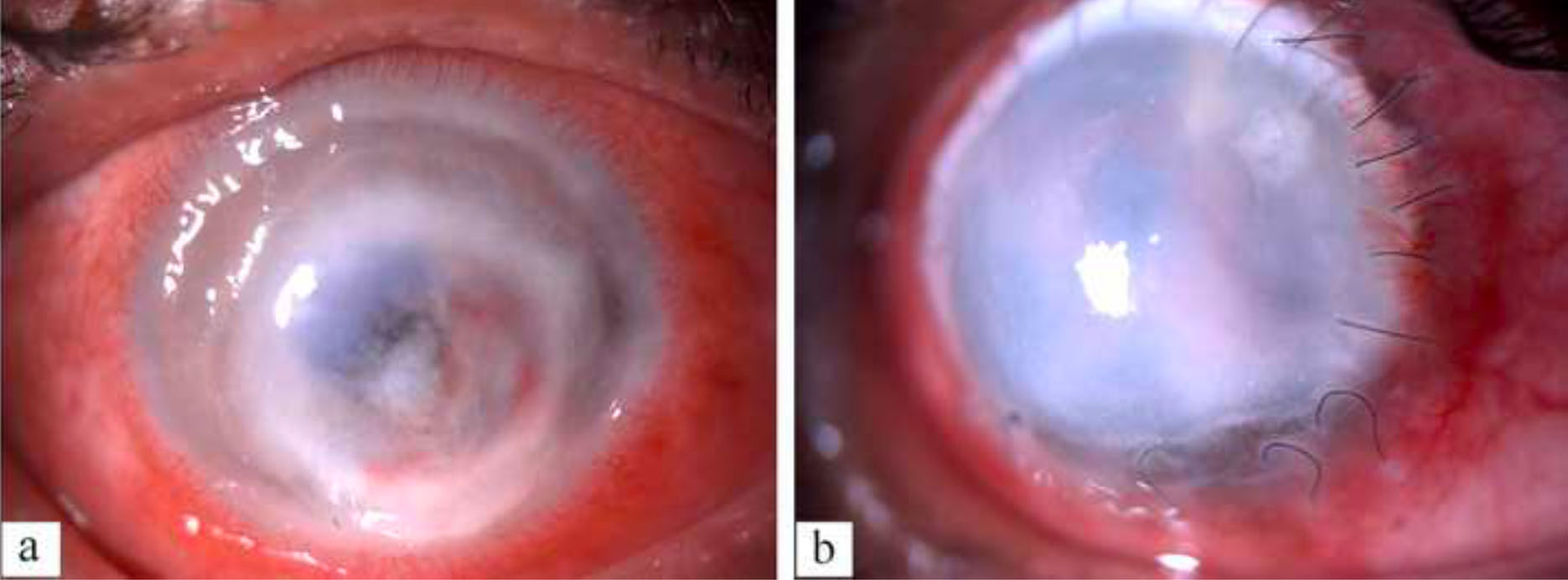
Figure 15 a- Digital slit lamp image of the patient depicting diffuse conjunctival congestion, superficial vascularization, total full thickness corneal infiltrate with 360 degree limbal infiltrate, paralimbal thinning with central impending perforation b- Digital slit lamp image of the patient depicting diffuse conjunctival congestion, total graft infiltrate, graft host junction melt from 1 to 7 o clock, loose sutures along with superficial vascularization
File history
Click on a date/time to view the file as it appeared at that time.
| Date/Time | Thumbnail | Dimensions | User | Comment | |
|---|---|---|---|---|---|
| current | 03:34, May 30, 2023 | 1,871 × 692 (123 KB) | Bharat.Gurnani (talk | contribs) |
You cannot overwrite this file.
File usage
The following page uses this file: