File:Esthesioneuroblastoma 20230620231423!Figure 2.jpg
From EyeWiki
No higher resolution available.
Esthesioneuroblastoma_20230620231423!Figure_2.jpg (438 × 242 pixels, file size: 65 KB, MIME type: image/jpeg)
Summary
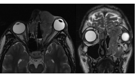
MRI showing a large left-sided nasal cavity mass with intracranial and orbital extension. There is a notable mass effect on the globe causing compression, proptosis and stretching of the optic nerve. Biopsy confirmed esthesioneuroblastoma. per https://eyewiki.org/w/index.php?title=File:Figure_2.jpg&oldid=56058
File history
Click on a date/time to view the file as it appeared at that time.
| Date/Time | Thumbnail | Dimensions | User | Comment | |
|---|---|---|---|---|---|
| current | 09:57, June 21, 2023 | | 438 × 242 (65 KB) | Tony.Ching.AAO (talk | contribs) | MRI showing a large left-sided nasal cavity mass with intracranial and orbital extension. There is a notable mass effect on the globe causing compression, proptosis and stretching of the optic nerve. Biopsy confirmed esthesioneuroblastoma. per https://eyewiki.org/w/index.php?title=File:Figure_2.jpg&oldid=56058 |
You cannot overwrite this file.
File usage
The following page uses this file: