File:Conjunctival amyloidosis.png
From EyeWiki
No higher resolution available.
Conjunctival_amyloidosis.png (513 × 382 pixels, file size: 398 KB, MIME type: image/png)
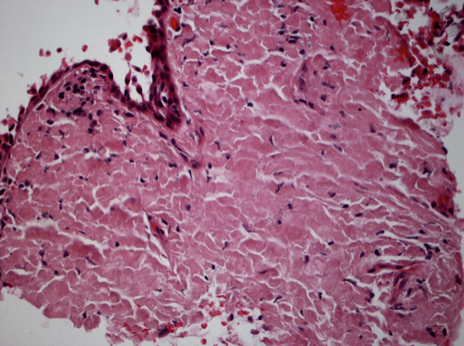
Figure 3: Conjunctival amyloidosis. Hematoxylin and eosin stain of conjunctival tissue biopsy. Extensive hyalinization and diffuse eosinophilic extracellular material consistent with amyloidosis. (Image courtesy of Paul J. Bryar, MD)
File history
Click on a date/time to view the file as it appeared at that time.
| Date/Time | Thumbnail | Dimensions | User | Comment | |
|---|---|---|---|---|---|
| current | 15:16, January 26, 2014 | | 513 × 382 (398 KB) | Frank.Hrisomalos (talk | contribs) | Figure 3: Conjunctival amyloidosis. Hematoxylin and eosin stain of conjunctival tissue biopsy. Extensive hyalinization and diffuse eosinophilic extracellular material consistent with amyloidosis. (Image courtesy of Paul J. Bryar, MD) |
You cannot overwrite this file.
File usage
The following page uses this file: