File:Chu OwlEye.png
From EyeWiki

Size of this preview: 800 × 393 pixels. Other resolution: 866 × 425 pixels.
Original file (866 × 425 pixels, file size: 460 KB, MIME type: image/png)
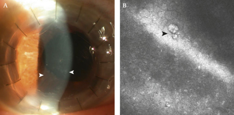
(A) Photograph of the right eye of the patient showing multiple coin-shaped keratic precipitates. (B) Confocal microscope of the right eye of the patient showing the owl's eye appearance in the corneal endothelium.
Chu HY, Sun CC, Chuang WY, et al. Cytomegalovirus associated corneal endotheliitis after penetrating keratoplasty in a patient with Fuchs corneal endothelial dystrophy. Br J Ophthalmol. 2012;96(2):300-301. doi:10.1136/bjo.2010.182378
File history
Click on a date/time to view the file as it appeared at that time.
| Date/Time | Thumbnail | Dimensions | User | Comment | |
|---|---|---|---|---|---|
| current | 12:21, March 16, 2021 | | 866 × 425 (460 KB) | Asim.farooq (talk | contribs) | (A) Photograph of the right eye of the patient showing multiple coin-shaped keratic precipitates. (B) Confocal microscope of the right eye of the patient showing the owl's eye appearance in the corneal endothelium. Chu HY, Sun CC, Chuang WY, et al. C... |
You cannot overwrite this file.
File usage
The following page uses this file: