File:Choroideremia Figure 5.png
From EyeWiki
No higher resolution available.
Choroideremia_Figure_5.png (512 × 384 pixels, file size: 268 KB, MIME type: image/png)
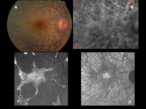
This image was originally published by BMC Ophthalmology. Authors: Maurizio BP, Pierluigi I, Stelios K, Stefano V, Marialucia C, Ilaria Z, Francesco B. Title: Retro-mode imaging and fundus autofluorescence with scanning laser ophthalmoscope of retinal dystrophies. This image is allowed to be used if properly cited, as per the Creative Commons Attribution License.© Parodi et al.; licensee BioMed Central Ltd. 2012
File history
Click on a date/time to view the file as it appeared at that time.
| Date/Time | Thumbnail | Dimensions | User | Comment | |
|---|---|---|---|---|---|
| current | 10:49, November 12, 2016 | | 512 × 384 (268 KB) | Peter.Bracha (talk | contribs) | This figure is a fundus photograph (A) and the associated fundus autofluorescence (C) of a patient with choroideremia. Notice the better characterization of diseased and healthy retina on fundus autofluorescence that is not as apparent as on the fundus... |
You cannot overwrite this file.
File usage
The following page uses this file: