File:Choroideremia Figure 4.jpg
From EyeWiki
No higher resolution available.
Choroideremia_Figure_4.jpg (643 × 532 pixels, file size: 133 KB, MIME type: image/jpeg)
Permission for use obtained from Director of Retina Image Bank and Members Section of the ASRS. This image was originally published in the Retina Image Bank. Author: Henry J. Kaplan, MD and Niloofar Piri, MD. Choroideremia. Retina Image Bank. March 29, 2013; Image Number 5375. © The American Society of Retina Specialists.
File history
Click on a date/time to view the file as it appeared at that time.
| Date/Time | Thumbnail | Dimensions | User | Comment | |
|---|---|---|---|---|---|
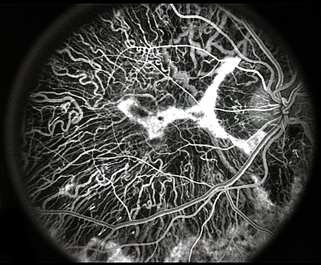
| current | 10:49, November 12, 2016 | | 643 × 532 (133 KB) | Peter.Bracha (talk | contribs) | This image is a fluorescein angiographic image in the early venous laminar phase demonstrating a irregular island of hyperfluorescence in the fovea surrounded by a generalized absence of choriocapillaris, retinal pigmented epithelium and retina. |
You cannot overwrite this file.
File usage
The following page uses this file: