File:Choroideremia Figure 3.jpg
From EyeWiki
No higher resolution available.
Choroideremia_Figure_3.jpg (600 × 450 pixels, file size: 70 KB, MIME type: image/jpeg)
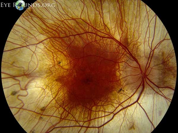
This image was originally published in the Ophthalmic Atlas Images by EyeRounds.org of The University of Iowa. Author: Jeffrey D. Welder, MD. Photographer: Toni Venckus, CRA. Title: Choroideremia. This image is licensed under the Creative Commons Attribution-NonCommercial-NoDerivs 3.0 Unported License. © of EyeRounds.org of The University of Iowa.Permissions obtained from executive director and editor of Eyerounds.org.
File history
Click on a date/time to view the file as it appeared at that time.
| Date/Time | Thumbnail | Dimensions | User | Comment | |
|---|---|---|---|---|---|
| current | 10:48, November 12, 2016 | | 600 × 450 (70 KB) | Peter.Bracha (talk | contribs) | This image is a fundus photograph of a patient with advanced choroideremia. Notice a relatively normal fovea surrounded by generalized atrophy of the neurosensory retina, retinal pigmented epithelium and choroid. |
You cannot overwrite this file.
File usage
The following page uses this file: