File:Choroidal pic.png

Original file (615 × 639 pixels, file size: 442 KB, MIME type: image/png)
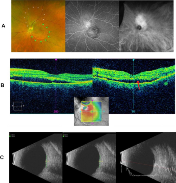
Choroidal Hemangioma: (A) shows fundus photo (left), FA(middle), and ICGA (right). FA revealed leakage from an ill-defined lesion superotemporal to the disc, while the ICGA showed diffuse and intense hypercyanescence of choroidal vessels, consistent with a choroidal hemangioma. (B) shows OCT of the same eye with foveal detachment and choroidal elevation (star). (C) B-scan ultrasonography and A-scan ultrasonography. A Masquerade Case: Choroidal Hemangioma Misdiagnosed As Central Serous Retinopathy © 2023 by Lai L, Javier T, Lee S, Gallemore RP. is licensed under Creative Commons Attribution – Non-Commercial (unported, v3.0) License.
File history
Click on a date/time to view the file as it appeared at that time.
| Date/Time | Thumbnail | Dimensions | User | Comment | |
|---|---|---|---|---|---|
| current | 10:38, November 23, 2023 | | 615 × 639 (442 KB) | Evan.Wotipka (talk | contribs) |
You cannot overwrite this file.
File usage
The following page uses this file: