File:Choroidal Macroaneurysm and Choroidal Macrovessels.jpg

Original file (688 × 935 pixels, file size: 626 KB, MIME type: image/jpeg)
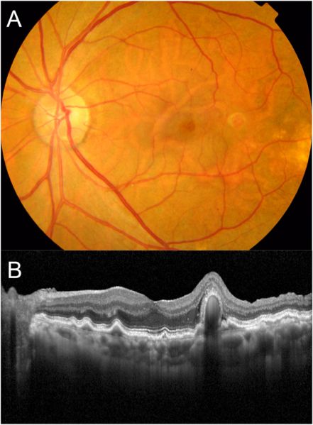
A: The color fundus photograph reveals an orange, protruding lesion within the curved segment of an anomalous choroidal macrovessel located in the temporal parafoveal region.
B: A horizontal SD-OCT scan through the fovea illustrates the presence of a protruding lesion in the sensory retina, accompanied by faint subretinal fluid.
Image source: Kinoshita T, Mori J, Imaizumi H. "Visual impairment associated with choroidal macroaneurysm in a patient with presumed anomalous short posterior ciliary artery." Am J Ophthalmol Case Rep. 2022 Nov 20;28:101755. doi: 10.1016/j.ajoc.2022.101755. PMID: 36439652; PMCID: PMC9694064. Licensed under Creative Commons Attribution (CC BY).
File history
Click on a date/time to view the file as it appeared at that time.
| Date/Time | Thumbnail | Dimensions | User | Comment | |
|---|---|---|---|---|---|
| current | 23:43, August 31, 2024 | | 688 × 935 (626 KB) | Joobin.Khadamy (talk | contribs) |
You cannot overwrite this file.
File usage
The following page uses this file: