File:Choroidal Folds Retinography.png
From EyeWiki
No higher resolution available.
Choroidal_Folds_Retinography.png (484 × 411 pixels, file size: 377 KB, MIME type: image/png)
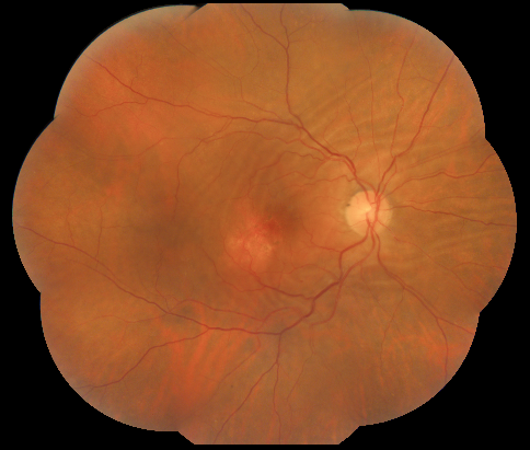
Choroidal folds in the posterior pole of the right eye associated with an area of atrophic and pigmentary changes (1 disc diameter in size) located inferotemporal to the fovea
File history
Click on a date/time to view the file as it appeared at that time.
| Date/Time | Thumbnail | Dimensions | User | Comment | |
|---|---|---|---|---|---|
| current | 11:15, November 9, 2020 | | 484 × 411 (377 KB) | Manuel.Marques (talk | contribs) | Choroidal folds in the posterior pole of the right eye associated with an area of atrophic and pigmentary changes (1 disc diameter in size) located inferotemporal to the fovea |
You cannot overwrite this file.
File usage
The following page uses this file: