File:Choroidal Folds Fluorescein Angiography.png
From EyeWiki

Size of this preview: 796 × 600 pixels. Other resolution: 831 × 626 pixels.
Original file (831 × 626 pixels, file size: 514 KB, MIME type: image/png)
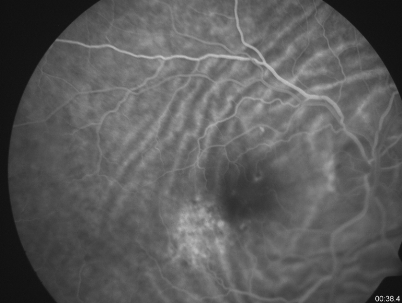
Fluorescein angiography shows an increased stippled hyperfluorescence with late staining corresponding to an area of pigmentary disturbance in the right eye. The folds are seen prominently due to their classic alternating hypo and hyperfluorescence
File history
Click on a date/time to view the file as it appeared at that time.
| Date/Time | Thumbnail | Dimensions | User | Comment | |
|---|---|---|---|---|---|
| current | 11:17, November 9, 2020 | | 831 × 626 (514 KB) | Manuel.Marques (talk | contribs) | Fluorescein angiography shows an increased stippled hyperfluorescence with late staining corresponding to an area of pigmentary disturbance in the right eye. The folds are seen prominently due to their classic alternating hypo and hyperfluorescence |
You cannot overwrite this file.
File usage
The following page uses this file: