File:Brain circuits for saccades.jpg
Original file (1,200 × 809 pixels, file size: 63 KB, MIME type: image/jpeg)
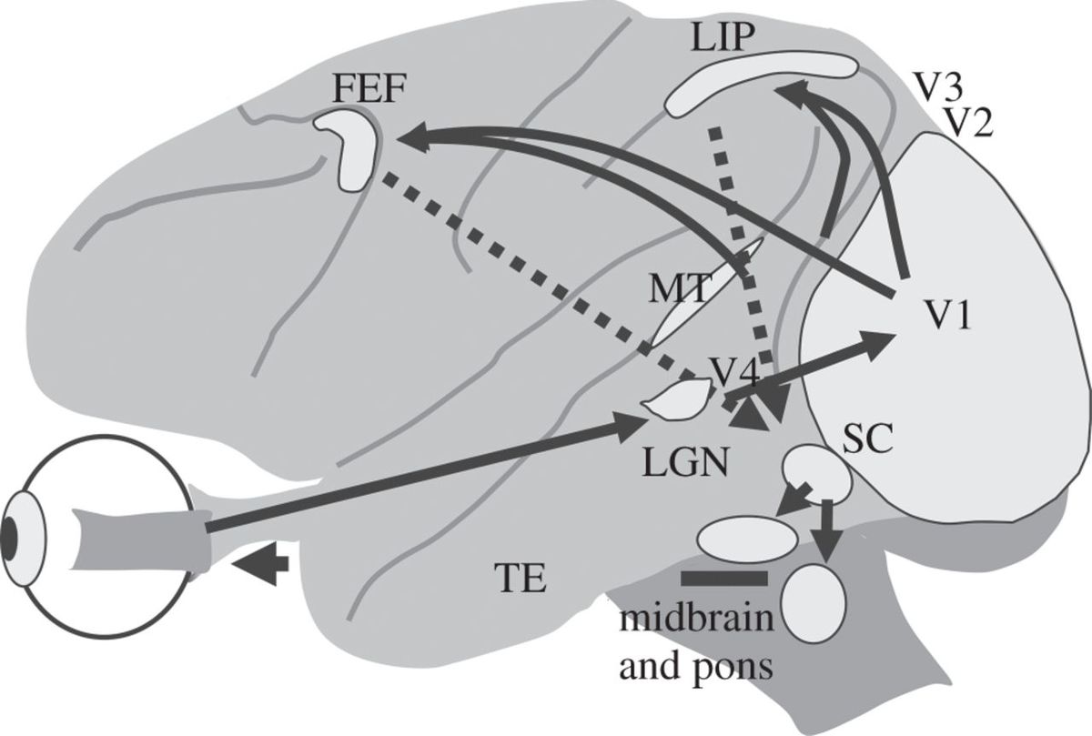
Description English: The brain circuits for visually guided saccades extend from cerebral cortex to the pons in the brain stem. This side view of the monkey brain shows that the circuit extends from retina to primary visual cortex (V1), then to extrastriate cortex, particularly to the lateral intraparietal (LIP) area and frontal eye field (FEF). From cortex, information reaches the superior colliculus (SC), and from there to brainstem oculomotor areas in midbrain and pons, and finally to the extraocular muscle motor neurons that project to the eye muscles to move the eye. This is a simplified brain circuit, and does not show a number of other circuits including those of the basal ganglia and the cerebellum. MT, middle temporal cortex; LGN, lateral geniculate nucleus; TE, anterior inferior temporal cortex. Date Published 3 August 2015 Source Robert H. Wurtz Using perturbations to identify the brain circuits underlying active vision Phil. Trans. R. Soc. B 2015 370 20140205; DOI: 10.1098/rstb.2014.0205. Published 3 August 2015 http://rstb.royalsocietypublishing.org/content/370/1677/20140205 Author Robert H. Wurtz
This file is licensed under the Creative Commons Attribution 4.0 International license.
File history
Click on a date/time to view the file as it appeared at that time.
| Date/Time | Thumbnail | Dimensions | User | Comment | |
|---|---|---|---|---|---|
| current | 09:16, April 15, 2019 | | 1,200 × 809 (63 KB) | Bayan.Al Othman (talk | contribs) | Description English: The brain circuits for visually guided saccades extend from cerebral cortex to the pons in the brain stem. This side view of the monkey brain shows that the circuit extends from retina to primary visual cortex (V1), then to extras... |
You cannot overwrite this file.
File usage
The following page uses this file: