File:BAMS composite EyeWiki.png
From EyeWiki

Size of this preview: 800 × 530 pixels. Other resolution: 2,471 × 1,636 pixels.
Original file (2,471 × 1,636 pixels, file size: 7.14 MB, MIME type: image/png)
Summary
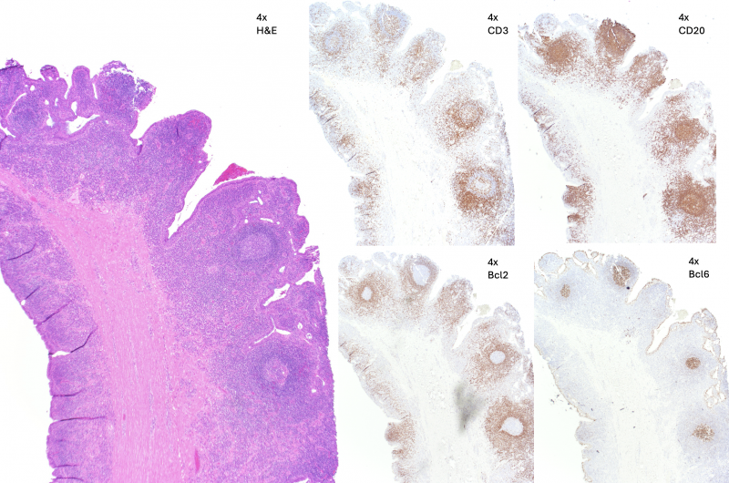
BAMS DCR specimen 4x view: Left: H&E stain highlighting dense lymphocytic infiltrate in a follicle formation with dense stroma consistent with a scar; Middle Left: CD3+ T cells highlighted in the normal expected location; Top Right: CD20+ B-cells in the germinal center as found normally; Bottom Left: Bcl2 + surrounding but not involving the germinal center, normal; Bottom Right: Bcl6+ in the germinal center, normal. Diagnosis of follicular lymphoid hyperplasia and scar.
File history
Click on a date/time to view the file as it appeared at that time.
| Date/Time | Thumbnail | Dimensions | User | Comment | |
|---|---|---|---|---|---|
| current | 17:17, September 12, 2024 | | 2,471 × 1,636 (7.14 MB) | Fabliha.Anbar (talk | contribs) | |
| 12:30, September 12, 2024 | 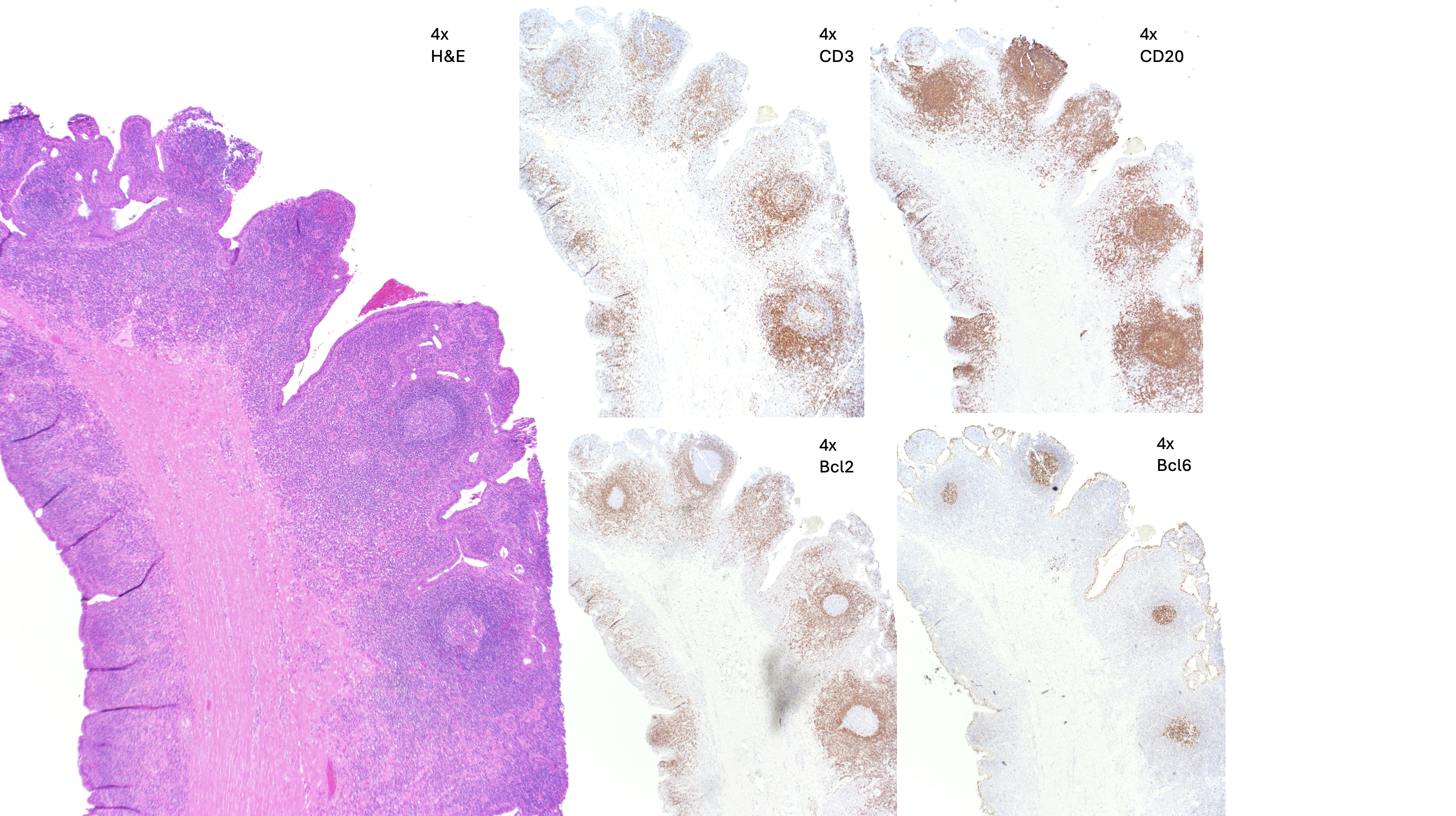 | 2,938 × 1,646 (7.66 MB) | Fabliha.Anbar (talk | contribs) | BAMS DCR specimen 4x view: Left: H&E stain highlighting dense lymphocytic infiltrate in a follicle formation with dense stroma consistent with a scar; Middle Left: CD3+ T cells highlighted in the normal expected location; Top Right: CD20+ B-cells in the germinal center as found normally; Bottom Left: Bcl2 + surrounding but not involving the germinal center, normal; Bottom Right: Bcl6+ in the germinal center, normal. Diagnosis of follicular lymphoid hyperplasia and scar. |
You cannot overwrite this file.
File usage
The following page uses this file: