File:ASRS-RIB-Image-28842.jpg

Original file (1,245 × 844 pixels, file size: 659 KB, MIME type: image/jpeg)
Summary
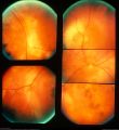
Choroidal Melanoma
By Karen Panzegrau
Photographer: Karen Panzegrau
Imaging device: Fundus Camera Optos
Description: Ultra-wide field Optos image of a 27-year-old male patient who presented with loss of vision for about 6-8 weeks. Previous choroidal nevus seen. Recommended annual monitoring. No exam since 10/2014. Brachytherapy vs enucleation was discussed. Brachytherapy was decided as treatment. Full metastatic workup is being performed.
Copyright notice: “This image was originally published in the Retina Image Bank® website. Karen Panzegrau. Choroidal Melanoma. Retina Image Bank. 2019; Image Number 3505. © the American Society of Retina Specialists."
File history
Click on a date/time to view the file as it appeared at that time.
| Date/Time | Thumbnail | Dimensions | User | Comment | |
|---|---|---|---|---|---|
| current | 17:40, December 8, 2021 |  | 1,245 × 844 (659 KB) | Drklai (talk | contribs) | |
| 17:34, December 8, 2021 |  | 1,889 × 2,058 (788 KB) | Drklai (talk | contribs) | Choroidal Melanoma By Karen Panzegrau Photographer Karen Panzegrau Imaging device Fundus camera Optos Description Ultra-wide field Optos image of a 27-year-old male patient who presented with loss of vision for about 6-8 weeks. Previous choroidal nevus seen. Recommended annual monitoring. No exam since 10/2014. Brachytherapy vs enucleation was discussed. Brachytherapy was decided as treatment. Full metastatic workup is being performed. Copyright notice: “This image was originally published in... |
You cannot overwrite this file.
File usage
The following 3 pages use this file:

