File:APMPPE.jpg
APMPPE.jpg (643 × 575 pixels, file size: 255 KB, MIME type: image/jpeg)
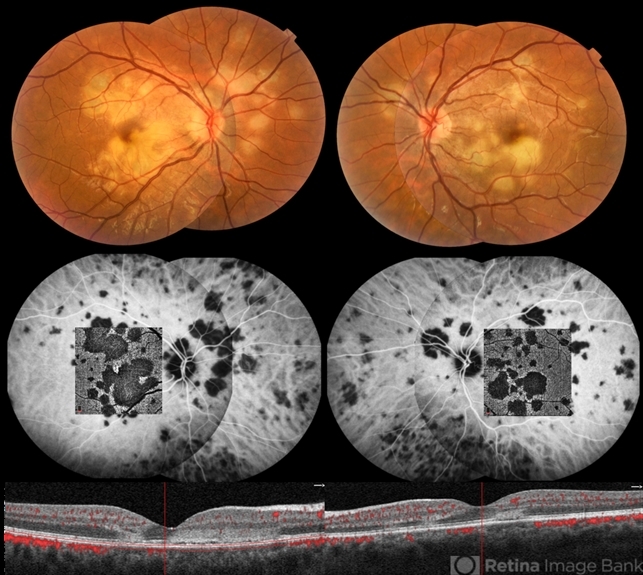
APMPPE - 27-year-old male with APMPPE, in the fundus photograph there are multiple yellow placoid lesions in the posterior pole of both eyes. ICGA revealed more lesions than those observed in fundoscopy. The OCTA segmented at the level of the choriocapillaris revealed areas of ischemia in close correspondence with the hypocyanescent lesions. The OCT with superimposed flow shows disruption and hyperreflectivity of the external retinal layers in the affected areas and absence of flow in the choriocapillaris underneath. This image was originally published in the Retina Image Bank® website. Claudia Farinha. Photographer Pedro Melo. Acute Posterior Multifocal Placoid Pigment Epitheliopathy. Retina Image Bank. 2019; 28757. © the American Society of Retina Specialists.
File history
Click on a date/time to view the file as it appeared at that time.
| Date/Time | Thumbnail | Dimensions | User | Comment | |
|---|---|---|---|---|---|
| current | 11:49, November 23, 2023 | | 643 × 575 (255 KB) | Evan.Wotipka (talk | contribs) |
You cannot overwrite this file.
File usage
The following page uses this file: