File:AA0 63833 Conjunctival–limbal autograft procedure.jpg
Original file (1,744 × 1,655 pixels, file size: 853 KB, MIME type: image/jpeg)
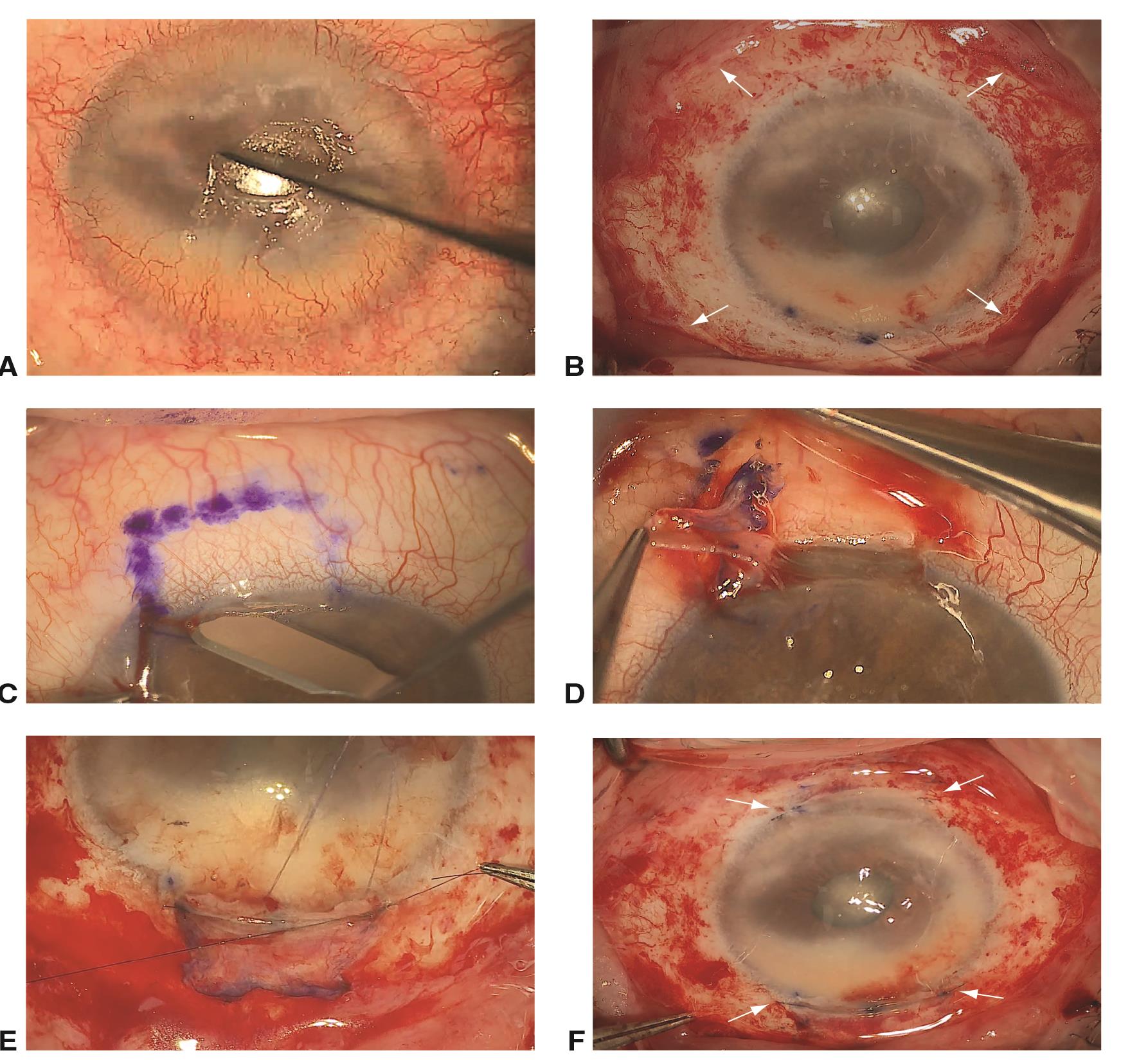
Conjunctival–limbal autograft procedure © 2023 American Academy of Ophthalmology
Conjunctival–limbal autograft procedure. A, Abnormal corneal epithelium and fibrovascular pannus are stripped by blunt dissection using a metal spatula or rounded blade and tissue forceps. B, The conjunctiva is undermined with blunt scissors, recessed 2–3 mm posterior to the limbus (arrows), and attached to the sclera with 10-0 polyglactin sutures or fibrin glue. A superior corneal traction suture is placed to facilitate exposure. C, Superior and inferior conjunctival–limbal grafts are delineated in the donor eye with a marking pen. A superficial incision is made within clear cornea with a crescent blade. D, The bulbar conjunctival part of the graft is undermined and thinly dissected from its limbal attachment. E, The limbal grafts are transferred to their corresponding sites in the recipient eye and secured with 10-0 nylon sutures at the corneal edge and 10-0 polyglactin sutures or fibrin glue at the conjunctival margin. F, Superior and inferior limbal grafts (arrows) at the conclusion of surgery.
File history
Click on a date/time to view the file as it appeared at that time.
| Date/Time | Thumbnail | Dimensions | User | Comment | |
|---|---|---|---|---|---|
| current | 20:26, March 8, 2023 | | 1,744 × 1,655 (853 KB) | Victoria.Chang (talk | contribs) |
You cannot overwrite this file.
File usage
The following page uses this file: