File:AA0 62636.jpg
From EyeWiki

Size of this preview: 800 × 571 pixels. Other resolution: 1,545 × 1,103 pixels.
Original file (1,545 × 1,103 pixels, file size: 166 KB, MIME type: image/jpeg)
Summary
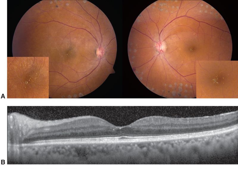
West African crystalline maculopathy. A, Fundus photographs of a patient from Nigeria with diabetes mellitus and previous panretinal photocoagulation scars. The distinct yellow-green refractile crystals in the fovea are better visualized in the magnified inset images. B, OCT image of the left eye shows the crystals located in the inner retina. Source: https://www.aao.org/image/west-african-crystalline-maculopathy Image License and Citation Guidelines: https://www.aao.org/asset.axd?id=76d72ec6-5f11-4f30-9242-a0c26b89f33e
File history
Click on a date/time to view the file as it appeared at that time.
| Date/Time | Thumbnail | Dimensions | User | Comment | |
|---|---|---|---|---|---|
| current | 09:11, May 13, 2021 | | 1,545 × 1,103 (166 KB) | Koushik.Tripathy (talk | contribs) | West African crystalline maculopathy. A, Fundus photographs of a patient from Nigeria with diabetes mellitus and previous panretinal photocoagulation scars. The distinct yellow-green refractile crystals in the fovea are better visualized in the magnified inset images. B, OCT image of the left eye shows the crystals located in the inner retina. Source: https://www.aao.org/image/west-african-crystalline-maculopathy Image License and Citation Guidelines: https://www.aao.org/asset.axd?id=76d72ec6... |
You cannot overwrite this file.
File usage
The following page uses this file: