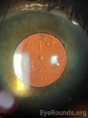
Elschnig Pearls
All content on Eyewiki is protected by copyright law and the Terms of Service. This content may not be reproduced, copied, or put into any artificial intelligence program, including large language and generative AI models, without permission from the Academy.
This article describes Elschnig Pearls and their management.
Disease Entity
Elschnig pearls are globular and semi-globular structures located on the posterior capsule that are believed to arise from regenerative posterior capsule opacification. They were initially identified by Hirshberg as a subtype of secondary cataract caused by the proliferation of equatorial lens fibres.1 Elschnig later delineated these formations from the alternative subtype of secondary cataract arising from posterior capsular fibrosis.1
Pathophysiology
Elschnig pearls are one of the various morphologies of regenerative posterior capsular opacification (PCO), a common long-term post-cataract surgery complication leading to decreased visual function after intraocular lens (IOL) implantation.2,3 The exact characterization of Elschnig pearls is quite unclear in the modern literature, which likely can be attributed to its rapid turnover in formation and drastic morphological changes in size and shape; however, various descriptions are proposed.3
Proposed Characterizations
Histologic observation has described Elschnig pearls as an attempted regenerative product of lens epithelial cells (LECs) migrating to proliferate between the IOL and posterior capsule.4 This phenomenon of LEC epithelial mesenchymal transition as a cellular substrate for PCO, and Elschnig pearls, has been indicated as a cytokine driven process.5 The eventual formation of syncytial PCO, then to Elschnig pearls, results in decreased visual acuity and contrast sensitivity loss.3
Elschnig pearls possess the ability to form extracellular vacuoles which appear as large, transparent spheres, similar to the ability of intact lenses to respond to exposure to extracellular fluid by increasing extracellular Na+/H2O transport, resulting in cyst-like cavity formation.6,7
Eslchnig pearls have also been described in rabbits as subcapsular epithelial cells originating from Soemmerring’s ring which escaped to migrate towards the center of the posterior capsule, however with transmission and EM scanning identifying different cell morphologies caught in between the IOL and posterior capsule.8-10
Other studies using transmission electron microscopy found no cell membrane remnants in Elschnig pearl collection, indicating them to be a biophysical product of a slow, lens fiber cell degradation process involving cell membrane ballooning instead;11 similar formations are also found in cataractous lenses as well.3 Their lack of an osmotic staining effect further suggests the presence of unsaturated lipid content.3
Their rapid size change, rate of appearance/disappearance, drastic size scale, low rate of fragmentation, and containment of highly refractive material compared to surrounding tissues further indicate for them to be cyst-like structures from lens fiber degeneration processes.3,12

Diagnosis
Elschnig pearls, like other forms of posterior capsular opacification (PCO), can be diagnosed by slit-lamp microscopy. They typically appear as clusters of pearls between the posterior chamber intraocular lens (PCIOL) and the posterior lens capsule.
Management
Like other manifestations of PCO, Elschnig pearls only require treatment if they become symptomatic and obstruct vision. Patients can be monitored clinically through vision screening, slit-lamp microscopy, or development of symptoms. If symptomatic, patients can undergo Nd:YAG laser capsulotomy which is the most common modality used for treating the fibrous and pearl forms of PCO.13 Another treatment option is surgical peeling and aspiration. Nd:YAG laser capsulotomy carries a higher risk of intraocular pressure (IOP) spikes and retinal detachment, while surgical peeling and aspiration is associated with the reappearance of pearls.13
Prognosis
Visual prognosis is very good with management. Collections of Elschnig pearls usually do not undergo spontaneous resolution. However, individual pearls have been reported to undergo rapid turnover consisting of progression and regression within days or weeks.3,14,15 Possible mechanisms for spontaneous resolution include apoptosis, falling of pearls into the vitreous humor through capsulotomy, and phagocytosis by macrophages.16
References
- Elschnig A. Klinisch-anatomischer Beitrag zur Kenntnis des Nachstares. Klin Monatsbl Augenheilkd. 1911;49:444–451.
- Neumayer T, Findl O, Buehl W, Sacu S, Menapace R, Georgopoulos M. Long-term changes in the morphology of posterior capsule opacification. J Cataract Refract Surg. 2005;31(11):2120-2128. doi:10.1016/j.jcrs.2005.04.038
- Findl O, Neumayer T, Hirnschall N, Buehl W. Natural course of Elschnig pearl formation and disappearance. Invest Ophthalmol Vis Sci. 2010;51(3):1547-1553. doi:10.1167/iovs.09-3989
- COWAN A, FRY WE. SECONDARY CATARACT: WITH PARTICULAR REFERENCE TO TRANSPARENT GLOBULAR BODIES. Arch Ophthalmol. 1937;18(1):12–22. doi:10.1001/archopht.1937.00850070024002
- Marcantonio JM, Vrensen GF. Cell biology of posterior capsular opacification. Eye (Lond). 1999;13 ( Pt 3b):484-488. doi:10.1038/eye.1999.126
- Fagerholm PP. On the formation of Elschnig's pearls. A tissue culture study of regenerating rat lens epithelium. Acta Ophthalmol (Copenh). 1980;58(6):963-970. doi:10.1111/j.1755-3768.1980.tb08323.x
- Sakuragawa M, Kuwabara T, Kinoshita JH, Fukui HN. Swelling of the lens fibers. Exp Eye Res. 1975;21(4):381-394. doi:10.1016/0014-4835(75)90048-2
- Kappelhof JP, Vrensen GF, Vester CA, Pameyer JH, de Jong PT, Willekens BL. The ring of Soemmerring in the rabbit: A scanning electron microscopic study. Graefes Arch Clin Exp Ophthalmol. 1985;223(3):111-120. doi:10.1007/BF02148886
- Kappelhof JP, Vrensen GF, de Jong PT, Pameyer J, Willekens B. An ultrastructural study of Elschnig's pearls in the pseudophakic eye. Am J Ophthalmol. 1986;101(1):58-69. doi:10.1016/0002-9394(86)90465-4
- Kappelhof JP, Vrensen GF, de Jong PT, Pameyer J, Willekens BL. The ring of Soemmerring in man: an ultrastructural study. Graefes Arch Clin Exp Ophthalmol. 1987;225(1):77-83. doi:10.1007/BF02155809
- Jongebloed WL, Kalicharan D, Los LI, van der Veen G, Worst JG. A combined scanning and transmission electronmicroscopic investigation of human (secondary) cataract material. Doc Ophthalmol. 1991;78(3-4):325-334. doi:10.1007/BF00165696
- Brown N. Visibility of transparent objects in the eye by retroillumination. Br J Ophthalmol. 1971;55(8):517-524. doi:10.1136/bjo.55.8.517
- Bhargava R, Kumar P, Sharma SK, Kaur A. A randomized controlled trial of peeling and aspiration of Elschnig pearls and neodymium: yttrium-aluminium-garnet laser capsulotomy. Int J Ophthalmol. 2015;8(3):590-596. Published 2015 Jun 18. doi:10.3980/j.issn.2222-3959.2015.03.28
- Nakashima Y, Yoshitomi F, Oshika T. Regression of Elschnig pearls on the posterior capsule in a pseudophakic eye. Arch Ophthalmol. 2002;120(3):397-398.
- Caballero A, Marín JM, Salinas M. Spontaneous regression of Elschnig pearl posterior capsule opacification. J Cataract Refract Surg. 2000;26(5):779-780. doi:10.1016/s0886-3350(00)00412-0
- Kurosaka D, Kato K, Kurosaka H, Yoshino M, Nakamura K, Negishi K. Elschnig pearl formation along the neodymium:YAG laser posterior capsulotomy margin. Long-term follow-up. J Cataract Refract Surg. 2002;28(10):1809-1813. doi:10.1016/s0886-3350(02)01222-1

