Corticosteroid-induced Glaucoma (Steroid Glaucoma) After Refractive Surgery
All content on Eyewiki is protected by copyright law and the Terms of Service. This content may not be reproduced, copied, or put into any artificial intelligence program, including large language and generative AI models, without permission from the Academy.
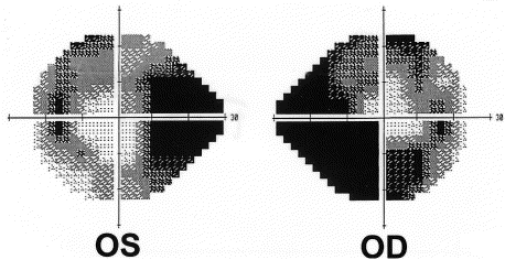
Steroid-induced glaucoma is a late-onset post-operative complication occurring as a result of normal post-operative regimen and/or following treatments for diffuse lamellar keratitis (DLK), another post-op complication. This secondary open-angle glaucoma is associated with high IOP and potential for permanent glaucomatous optic neuropathy. Detection of the condition may be difficult due to falsely-low intraocular pressure readings associated with thin corneas and intra-stromal fluid. It is imperative to make an early diagnosis as continued treatment with corticosteroids can produce serious and irreversible visual loss. Simple discontinuation or tapering of corticosteroids often leads to cessation of symptoms; however, some eyes can have a peristenly higher IOP compared to baseline.
Clinical Features
- Worsening visual acuity and loss of visual field
- Increased measured intraocular pressure
- Worsening symptoms with corticosteroid
- Interface fluid on slit-lamp biomicroscopy, though not seen in all cases
- Lack of mononuclear cells and granulocyte-like cells by confocal microscopy
- Minimal inflammatory cells
- Cystic corneal edema beyond flap margin
- Thickened flap
- Central steepening corneal topography—most likely due to interface fluid pushing central cornea anteriorly
- Intra-ocular pressure hypotony of less than 9 mm by applanation tonometry; hypertony on flap periphery by Tono-Pen [1].
- Sometimes associated with late-onset DLK occurring more than 10 days following surgery
- Occurrence at approximately two weeks after administration of steroids
Didactic Case
Patient: 30 yo male who received bilateral LASIK [2]
| Preoperative manifest refraction | -10.25 - 0.75 x 007 right eye
-12 - 2.00 x 140 left eye Best corrected visual acuity 20/30 in each eye | |
|---|---|---|
| 1 week post- operative (PO) | Condition | Mild irritation in left eye, thus diagnosed with diffuse lamellar keratitis |
| Treatment | Topical prednisolone acetate every 2 hrs, both eyes | |
| 3 weeks PO | Condition |
|
| Treatment | Brimonidine and timolol (both for treating ocular hypertension), oral methyprednisolone and topical prednisolone every hour in each eye to treat inflammation | |
| 4 weeks PO | Condition | Visual acuity diminish to 20/200 in left eye |
| Treatment | Start oral prednisone once daily, continue hourly topical prednisolone to treat DLK more aggressively | |
| 5 weeks PO | Condition | Left eye visual acuity: 20/400 |
| Treatment | Oral steroids tapered, topical steroids hourly in both eyes | |
| 7 weeks PO | Condition |
|
| Treatment | Stop oral and topical steroid medications, and continue IOP-reducing medication (brimonidine and timolol), and start acetazolamide, anti-glaucoma drug | |
| 8 weeks PO | Condition | Visual acuity of 20/40 right eye and 20/50 left eye |
| Treatment |
|

Case Summary
Patient with diagnosis of DLK one week following refractive surgery is treated for associated inflammation with both topical and oral steroid. Symptoms worsen, and severely elevated IOP in both eyes is detected 6 weeks following initial DLK diagnosis. When IOP and associated glaucoma are treated by stopping steroids and starting anti-glaucoma medication, condition including visual acuity markedly and rapidly improves.
Theories of Mechanism
The clinical element that often obscures diagnosis of steroid-induced glaucoma and rise in IOP is the falsely low IOP measurements in affected patients. Applanation tonometry assumes a corneal thickness of 520 µm, and the invariable negative deviation from the normal thickness as a result of refractive surgeries lead to lower IOP readings. It is estimated that with central corneal thickness is 450 µm, IOP is underestimated by 5.2 mm Hg [3]. Falsely low IOP has also been attributed to intra-stromal fluid collecting just beneath the flap [4][2]
It is generally agreed that the IOP elevation due to steroid administration results from reduction in facility of aqueous outflow [5]. Several theories have been proposed to explain how corticosteroid manifests its effect on the outflow.
- Steroid alters trabecular meshwork cell morphology by increasing nuclear transport of the human glucocorticoid receptor GRbeta [6].
- Corticosteroids increase expression of extracellular matrix protein fibronectin, polymerized glycosaminoglycans, and elastin. Elevated amounts of named substances thus lead to their accumulation in the trabecular meshwork, obstructing the outflow pathway [7].
- The endothelial cells of the trabecular meshwork are phagocytic and can thus remove and destroy debris entering the meshwork from the anterior chamber. Corticosteroids are known to suppress phagocytic activity, and supression of phagocytosis mayallow debris in the aqueous humor to accumulate and act as a barrier to outflow [8].
- Corticosteroids cause physical obstruction of the trabecular meshwork with pigmented, crystalline steroid particles [9].
Risk Factors
As with common steroid-induced glaucoma, individuals with pre-existing chronic open-angle glaucoma and family history of the disease are at greater risk compared to individuals without such history. Other risk factors are high myopia, previous steroid response, African-American ethnicity, age, history of connective tissue disease, especially rheumatoid arthritis [10], and Type 1 diabetes mellitus. Although there are case reports of glaucomatous progression following refractive surgery, several well-designed studies demonstrate no significant change in retinal nerve fiber analysis after either either LASIK or LASEK. [1]
Prevalence
The incidence of increased IOP after surface ablation has been reported to range from 11% to 25% of patients [11]. The incidence is higher can be as high as 25% in patients using dexamethasone or similar strong corticosteroids compared to patients using fluorometholone (incidence of 1.5-3% of patients).
Management
Treating glaucoma begins with topical medication, frequent pressure checks and monitoring corneal topography to track improvement and management. Management is similar to that of regular glaucoma with minor differences. IOP and corneal topography should be monitored regularly to track improvement and management.
- Stop steroids. This is often all that is required [5][3]
- Start anti-glaucoma meds, such as systemic acetazolamide and topical timolol
- Adrenergic agonists
- Beta-blockers (e.g., timolol)
- Sympathomimetics
- Carbonic anhydrase inhibitors (e.g., acetazolamide)
- Miotic agents o prostaglandin analogs
- Surgical
- Excision of depot steroid
- Laser trabeculoplasty
- Trabeculectomy
- Glaucoma Drainage Devices
- Cyclophotocoagulation
Prevention
It is urged that patients receiving steroid therapy, either systemically or locally to the eye, be examined periodically for possible elevations in intraocular pressure. Patients who have a family history or previous diagnosis of open-angle glaucoma should be observed more frequently [5]. Refractive surgeries should not be considered when IOP is poorly controlled. Provider must recognize that the interface fluid characteristic of post-refractive surgery steroid-induced glaucoma leads to inaccurately low central applanation tonometry measurements and obscures the diagnosis of steroid-induced glaucoma. Measurements of central cornea should be confirmed with those obtained from the cornea peripheral—preferably temporal rather than nasal--to the flap using Tono-Pen, pneumotonometery, or applanation tonometer[2][4][7].
Additional Resources
References
- ↑ Jump up to: 1.0 1.1 Sharma N, Sony P, Gupta A, Vajpayee RB. Effect of laser in situ keratomileusis and laser-assisted subepithelial keratectomy on retinal nerve fiber layer analysis. J Cataract Refract Surg. 2006; 32:446-450.
- ↑ Jump up to: 2.0 2.1 2.2 2.3 Hamilton DR, Manche EE, Rich LF, Maloney RK. Steroidinduced glaucoma after laser in situ keratomileusis associated with interface fluid. Ophthalmology 2002;109:659–65.
- ↑ Jump up to: 3.0 3.1 Allingham RR, Damji KF, Freedman S, Moroi SE, Shafranov G. Shields' Textbook of Glaucoma. 5th Edition. Lippincott Williams & Wilkins. 2004
- ↑ Jump up to: 4.0 4.1 Cheng AC, Law RW, Young AL, Lam DS. In vivo confocal microscopic findings in patients with steroid-induced glaucoma after LASIK. Ophthalmology. 2004 Apr;111(4):768-74.
- ↑ Jump up to: 5.0 5.1 5.2 Jones R, Rhee DJ. Corticosteroid-induced ocular hypertension and glaucoma: a brief review and update of the literature. Curr Opin Ophthalmol. 2006;17(2):163-167.
- ↑ Wordinger RJ, Clark AF. Effects of glucocorticoids on the trabecular meshwork: towards a better understanding of glaucoma. Prog Retin Eye Res. 1999;18(5):629-667.
- ↑ Jump up to: 7.0 7.1 Steeley HT, Browder SL, Julian MB, et al. The effects of dexamethasone on fibronectin expression in cultured human trabecular meshwork cells. Invest Ophthalmol Vis Sci. 1992;33(7):2242-2250.
- ↑ Bill A. The drainage of aqueous humor [editorial]. Invest Ophthalmol. 1975;14(1):1-3.
- ↑ Im L, Allingham RR, Singh I, Stinnett S, Fekrat S. A prospective study of early intraocular pressure changes after a single intravitreal triamcinolone injection. J Glaucoma. 2008 Mar;17(2):128-32.
- ↑ Rhee DJ, Gedde S. "Glaucoma, Drug-Induced" May 18, 2009 2009. Web. <http://emedicine.medscape.com/article/1205298-overview>.
- ↑ Rapuano CJ. 2008 - 2009 Basic and Clinical Science Course (BCSC) Section 13 Refractive Surgery. American Academy of Ophthalmology. San Francisco, California. 2008.
- Becker B, Bernstein HN, Mills DW. Steroid-Induced Elevationof Intraocular Pressure. Archives of Ophthalmology. 1963;70(1):15-18.

