Congenital Simple Hamartoma of the Retinal Pigment Epithelium (CSHRPE)
All content on Eyewiki is protected by copyright law and the Terms of Service. This content may not be reproduced, copied, or put into any artificial intelligence program, including large language and generative AI models, without permission from the Academy.

Disease Entity
Congenital Simple Hamartoma of the Retinal Pigment Epithelium (CSHRPE) is indeed a rare intraocular condition characterized by focal, nodular lesions, often located near the fovea. Due to its rarity and incidental presentations in patients seeking care for other symptoms, diagnosis can be challenging, often requiring advanced imaging techniques such as Optical Coherence Tomography (OCT), OCT Angiography (OCTA), Fluorescein Angiography (FA), and Indocyanine Green Angiography (ICG).
While CSHRPE is generally considered benign, it can lead to visual impairment, particularly if associated with complications such as macular edema or vitreoretinal traction. In such cases, interventions like vitrectomy or anti-VEGF therapy may be considered to preserve or improve vision.
Given the limited understanding of its etiology and potential impact on retinal function, further research into CSHRPE is essential. Future studies should aim to elucidate its underlying causes and better understand its effects on retinal structure and function, ultimately contributing to improved diagnosis, management, and outcomes for affected individuals.
Overview
Pigmented tumors situated in the posterior segment of the eye display a wide range of origins and characteristics. Originating from either the retinal pigment epithelium (RPE) or the choroidal melanocytes, these tumors are classified as either benign or malignant. One such benign tumor is congenital simple hamartoma of the retinal pigment epithelium (CSHRPE), a rare hyperpigmented lesion, either solitary or multifocal, located near the fovea.
Disease
Congenital Simple Hamartoma of the Retinal Pigment Epithelium (CSHRPE) is a rare intraocular condition characterized by single or multiple focal, nodular lesions often found juxtafovea. Diagnosis can be challenging due to its varied presentations and similarities to other retinal conditions, but advanced imaging techniques like OCT, OCTA, FA, and ICG play a crucial role. While CSHRPE is typically benign, it can cause visual impairment if associated with macular edema or vitreoretinal traction, prompting consideration for interventions such as vitrectomy or anti-VEGF therapy. Future research should focus on elucidating its etiology, and understanding its functional impact on the retina.[1]
Etiology
CSHRPE is believed to have a congenital origin, with its pathogenesis proposed to involve disturbances in the signaling pathways as RPE cells migrate during embryonic development.[2]
Risk Factors
The specific risk factors for Congenital Simple Hamartoma of the Retinal Pigment Epithelium (CSHRPE) are not yet fully understood. However, some hypotheses suggest that intrauterine toxin exposure or viral infections during pregnancy may potentially contribute to its development. Further research is needed to elucidate the exact risk factors and underlying mechanisms involved in the pathogenesis of CSHRPE.[3]
Diagnosis
Diagnosing CSHRPE can be challenging due to its benign nature and lack of symptoms. Advanced imaging modalities play a crucial role in differentiation, offering valuable insights into the structural aspects of CSHRPE.
Clinical Features
- Appearance: CSHRPE typically presents as a solitary or multiple nodular hyperpigmented lesion.[3]
- Location: These lesions are usually found juxtafovea.
- Visual Acuity: Patients generally maintain normal visual acuity unless the lesion encroaches upon or involves the fovea, which may lead to visual impairment.
Imaging Modalities
The clinical findings and imaging modalities described in the case presentation of Congenital Simple Hamartoma of the Retinal Pigment Epithelium (CSHRPE) can be summarized as follows:
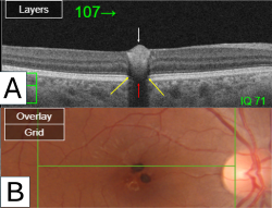
- Fundus photo: may show single or multiple pigmented nodules, intraretinal yellow materials, and radial retinal wrinkles.
- Red-free (RF) Imaging, Infrared (IR) Imaging, Enhanced IR Imaging, and Shadowgram: Hyperpigmented lesions appeared dark in all these imaging modalities, RF accentuates radial folds on the internal surface of the retina.
- Optical Coherence Tomography (OCT): Shows hyperreflective lesions protruding toward the vitreous cavity, accompanied by posterior shadowing, resulting in abrupt hyporeflectivity resembling pseudodisruption of the photoreceptor and RPE layer.
- Three-dimensional (3D) Rendered OCT: Enhances the visualization of pigmented nodules protruding toward the vitreous, along with radial wrinkles in the internal limiting membrane.
- OCT Angiography (OCTA): may show high signal intensity and flow at nodules in the superficial capillary plexus slab (SCP).
- Fluorescein Angiography (FA): shows blockage at the site of hyperpigmented lesions with late ring-form hyperfluorescence staining between nodules.
- Indocyanine Green Angiography (ICGA): Demonstrates blockage at the site of hyperpigmented lesions with no evidence of leakage or staining in the late phase.
These findings collectively contribute to the diagnosis and understanding of CSHRPE, providing valuable insights into its clinical presentation and imaging characteristics.[3]
Differential Diagnosis
The differential diagnosis for Congenital Simple Hamartoma of the Retinal Pigment Epithelium (CSHRPE) includes several conditions:
- Congenital Hypertrophy of the Retinal Pigment Epithelium (CHRPE): Similar in appearance to CSHRPE, CHRPE presents as a solitary or multiple darkly pigmented lesions often found in the midperiphery of the retina.
- Combined Hamartoma of the Retina and Retinal Pigment Epithelium (RPE): This condition involves both retinal and RPE elements, characterized by a gray, elevated mass with retinal vessels coursing over its surface.
- Adenoma or Adenocarcinoma of the RPE: Neoplasms originating from the RPE can mimic the appearance of CSHRPE, often presenting as elevated, pigmented lesions with variable vascularity and sometimes associated subretinal fluid or exudates.
- Retinal Invasion from Underlying Choroidal Nevus or Melanoma: Choroidal lesions such as nevi or melanomas can extend into the retina, presenting as elevated pigmented lesions with associated subretinal fluid or exudates.
- Intraretinal Foreign Body: Presence of a foreign body within the retina can mimic the appearance of a pigmented retinal lesion, particularly if there is associated inflammation or hemorrhage.
Each of these conditions presents unique clinical and imaging features that help differentiate them from CSHRPE. Accurate diagnosis relies on careful evaluation of clinical findings and imaging characteristics, often necessitating a combination of modalities for definitive diagnosis and appropriate management.[1]
Management
While CSHRPE typically demonstrates a benign course, interventions such as anti-vascular endothelial growth factor (anti-VEGF) injections or vitrectomy may be performed in cases where complications arise.[3]
Complications
Complications associated with CSHRPE may include compromised vision, macular edema, vitreomacular traction, epiretinal membrane, and macular hole.[3]
Prognosis
The prognosis for CSHRPE is generally favorable, with most lesions remaining stable or exhibiting slow or no growth over time.[3]
Additional Resources
Include relevant articles, books, or websites for further reading on CSHRPE.
- Laqua H: Tumors and tumor-like lesions of the retinal pigment epithelium. Ophthalmologica. 1981, 183:34-8. 10.1159/000309131
- Gass JD: Focal congenital anomalies of the retinal pigment epithelium. Eye. 1989, 3 ( Pt 1):1-18. 10.1038/eye.1989.2
- Rodrigues MW, Cavallini DB, Dalloul C, et al.: Retinal sensitivity and photoreceptor arrangement changes secondary to congenital simple hamartoma of retinal pigment epithelium. Int J Retina Vitreous. 2019, 5:5. 10.1186/s40942-018-0154-7
- Shields JA, Shields CL: Tumors and Related Lesions of the Pigmented Epithelium. The Asia-Pacific Journal of Ophthalmology. 2017, 6:215. 10.22608/APO.201705
- Ito Y, Ohji M: Long-Term Follow-Up of Congenital Simple Hamartoma of the Retinal Pigment Epithelium: A Case Report. Case Rep Ophthalmol. 2018, 9:215-20. 10.1159/000487631
- Bach A, Gold AS, Villegas VM, et al.: Simple hamartoma of the retinal pigment epithelium with macular edema. Optom Vis Sci. 2015, 92:48-50. 10.1097/OPX.0000000000000534
- Stavrakas P, Vachtsevanos A, Karakosta E, et al.: Full-thickness macular hole associated with congenital simple hamartoma of retinal pigment epithelium (CSHRPE). Int Ophthalmol. 2018, 38:2179-82. 10.1007/s10792-017-0676-2
- Naseripour M, Hemmati S, Aghili SS, et al.: Congenital simple hamartoma of the retinal pigment epithelium: 4 cases with multimodal imaging. Ophthalmic Genet. 2024, 45:78-83. 10.1080/13816810.2023.2206889
- Shukla D, Ambatkar S, Jethani J, et al.: Optical coherence tomography in presumed congenital simple hamartoma of retinal pigment epithelium. Am J Ophthalmol. 2005, 139:945-7. 10.1016/j.ajo.2004.11.037
- Heo WJ, Park DH, Shin JP: A Case of Congenital Simple Hamartoma of the Retinal Pigment Epithelium and Coats’ Disease in the Same Eye. Korean J Ophthalmol. 2015, 29:282-3. 10.3341/kjo.2015.29.4.282
- Souissi K, El Afrit MA, Kraiem A: Congenital retinal arterial macrovessel and congenital hamartoma of the retinal pigment epithelium. J Pediatr Ophthalmol Strabismus. 2006, 43:181-2. 10.3928/01913913-20060301-10
- Badawi AH, Magliyah M, Allam K, et al.: Foveal Congenital Simple Hamartoma of Retinal Pigment Epithelium: A Report of Two Cases. Middle East Afr J Ophthalmol. 2020, 27:128-30. 10.4103/meajo.MEAJO_177_20
- Tripathy K, Bandyopadhyay G, Basu K, et al.: Congenital simple hamartoma of the retinal pigment epithelium with depigmentation at the margin in an Indian female. GMS Ophthalmol Cases. 2019, 9:23. 10.3205/oc000112
- Shields CL, Manalac J, Das C, et al.: Review of spectral domain-enhanced depth imaging optical coherence tomography of tumors of the retina and retinal pigment epithelium in children and adults. Indian J Ophthalmol. 2015, 63:128-32. 10.4103/0301-4738.154384
- Arjmand P, Elimimian EB, Say EAT, et al.: OPTICAL COHERENCE TOMOGRAPHY ANGIOGRAPHY OF CONGENITAL SIMPLE HAMARTOMA OF THE RETINAL PIGMENT EPITHELIUM. Retin Cases Brief Rep. 2019, 13:357-60. 10.1097/ICB.0000000000000596
- Teke MY, Ozdal PÇ, Batioglu F, et al.: Congenital simple hamartoma of retinal pigment epithelium: clinical and imaging findings. Case Rep Ophthalmol Med. 2012, 2012:654502. 10.1155/2012/654502
References
Provide citations for all sources referenced in the article.
- ↑ Jump up to: 1.0 1.1 Shields CL, Shields JA, Marr BP, et al.: Congenital simple hamartoma of the retinal pigment epithelium: a study of five cases. Ophthalmology. 2003, 110:1005-11. 10.1016/S0161-6420(03)00087-3
- ↑ Barnes AC, Goldman DR, Laver NV, et al.: Congenital simple hamartoma of the retinal pigment epithelium: clinical, optical coherence tomography, and histopathological correlation. Eye. 2014, 28:765-6. 10.1038/eye.2014.49
- ↑ Jump up to: 3.0 3.1 3.2 3.3 3.4 3.5 Khadamy J, Elmi Sadr N (May 21, 2024) Multifocal Congenital Simple Hamartoma of the Retinal Pigment Epithelium: A Multimodal Imaging Case Study. Cureus 16(5): e60755. doi:10.7759/cureus.60755

