Cerebro-Oculo-Facial-Skeletal Syndrome
All content on Eyewiki is protected by copyright law and the Terms of Service. This content may not be reproduced, copied, or put into any artificial intelligence program, including large language and generative AI models, without permission from the Academy.
Cranio-oculo-facio-skeletal syndrome (COFS) is a rare congenital autosomal recessive disorder impairing the development of the brain, the head, the eyes, the limbs, and the face.[1]
Disease Entity
COFS is caused by a defect in the nucleotide excision repair pathway and can be diagnosed prenatally via imaging and genetic testing.[1] Based on a case report by Pena et al., most individuals do not survive beyond 30 months.[2] Ocular manifestations can include blepharophimosis, microphthalmia, nystagmus, cataracts, and hypertelorism.[3] Ruling out COFS is critical to guiding management with regards to ocular outcomes and procedures in other conditions, most notably cataract surgery.
Disease Epidemiology
Only 14 cases of COFS were documented between 1974 and 2010.[3] The only documented case of COFS since 2010 was reported in 2021 by Sirchia et al.[1]
Etiology
COFS is a congenital autosomal recessive disorder stems from a mutation in the transcription coupled nucleotide excision repair pathway.[3] Possible affected genes include the Cockayne syndrome gene (CSB) and the Xeroderma Pigmentosum genes (XPD, XPG, ERCCI).
Risk Factors
Frequent consanguineous marriages are a significant risk factor for COFS.[4] Microarray analysis and targeted molecular testing for mutations in the following genes can help determine a couple’s risk for conceiving a child with COFS:
- 46 nucleotide excision repair genes, most notably:[1]
- ERCC1
- ERCC2
- ERCC5
- ERCC6
- KIAA1109
- PHGDH
- FKTN
Pathophysiology
Defects in the Nucleotide Excision Repair pathway lead to the accumulation of genetic mutations culminating in the disease presentation of COFS.[1]
Diagnosis
- Clinical criteria for diagnosis:[5]
- Microcephaly
- Congenital cataracts
- Microphthalmia
- Multiple joint contractures
- Growth failure and developmental delay
- Facial dysmorphisms
- Prominent nasal root
- Overhanging upper lip
- Genetic Criteria for diagnosis:[5]
- DNA repair defect in Nucleotide Excision Repair pathway
Physical Examination
- Craniofacially, the disorder can manifest with:[3]
- Microcephaly
- Micrognathia
- Small mouth
- Cleft palate
- High arched palate
- Short neck
- Neurologically, COFS can manifest with the following findings:[3]
- Hyporeflexia or areflexia throughout throughout the body
- Sensorineural hearing loss
- Impaired cognitive development
- Other musculoskeletal manifestations of COFS include:[3]
- Arthrogryposis
- Flexion contractures, especially in the elbows and knees
- Hypotonia
- Syndactyly
- Rocker bottom feet
- Osteoporosis
- Arthrogryposis
Ocular Findings
- Blepharophimosis[3]
- Microphthalmia
- Nystagmus
- Cataracts
- Hypertelorism
Laboratory Testing & Imaging
Genetic Testing:[1]
- Microarray analysis
- NGS panels
- Whole Exome Sequencing
- Whole Genome Sequencing
Imaging:
- Radiography[6]
- Generalized under-mineralization of the skeleton
- Microcephaly
- CT[7]
- Intracranial calcifications
- MRI[7]
- progressive demyelination of the brain
- Ventriculomegaly
- Cerebellar dysplasia
- partial or complete degeneration of the corpus callosum
- Ultrasound[1]
- Clenched hand and abducted fingers
- Rockerbottom feet
- Bilateral microphthalmia with cataract formation
- Micrognathia with low set ears
Differential diagnosis
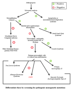
The differential diagnosis of conditions that mimic COFS is important to guiding management decisions in both COFS and in other conditions. A key example for many of the conditions included in the differential is determining the utility of cataract surgery. For instance, a part of the recommended management of Cockayne syndrome, a more common condition that presents similarly to COFS, is to conduct cataract surgery soon after the detection of cataracts. This is because, when performed before the onset of retinal dystrophy which develops later on, visual outcomes improve.[8] For these patients, cataract surgery is a meaningful intervention that can help them maximize their limited life span of 12 years. In contrast, cataract surgery is not performed in COFS patients, who live to about 30 months in most cases.[9]
- Differential Diagnosis includes:
- Cockayne Syndrome[7]
- Alkuraya-Kucinskas Syndrome[1]
- Neu-Laxova Syndrome[3][1]
- Muscular dystrophy-dystroglycanopathy type A4[1]
- Primary microcephaly-10[1]
- Costello Syndrome[1]
- Smith-Lemli-Opitz Syndrome[1]
- Micro Syndrome (Warburg-Microsyndrome)[3]
- Martsolf Syndrome[3]
- Cataract, Microcephaly, Failure to Thrive, Kyphoscoliosis Syndrome (CAMFAK)[3]
Management & Outcomes
Ophthalmic findings can be managed per the usual protocols.
According to a case report by Lowry et al., failure to thrive due to feeding problems along with recurrent aspiration pneumonia led to death prior to 30 months of age in 8 out of 10 patients.[2]
References
- ↑ Jump up to: 1.00 1.01 1.02 1.03 1.04 1.05 1.06 1.07 1.08 1.09 1.10 1.11 1.12 Sirchia, F., Fantasia, I., Feresin, A., Giorgio, E., Faletra, F., Mordeglia, D., Barbieri, M., Guida, V., De Luca, A., & Stampalija, T. (2021). Prenatal findings of cataract and arthrogryposis: recurrence of cerebro-oculo-facio-skeletal syndrome and review of differential diagnosis. BMC medical genomics, 14(1), 89. https://doi.org/10.1186/s12920-021-00939-6
- ↑ Jump up to: 2.0 2.1 Pena, S. D., Evans, J., & Hunter, A. G. (1978). COFS syndrome revisited. Birth defects original article series, 14(6B), 205–213.
- ↑ Jump up to: 3.00 3.01 3.02 3.03 3.04 3.05 3.06 3.07 3.08 3.09 3.10 Suzumura, H., & Arisaka, O. (2010). Cerebro-oculo-facio-skeletal syndrome. Advances in experimental medicine and biology, 685, 210–214. https://doi.org/10.1007/978-1-4419-6448-9_19
- ↑ Graham Jr, J. M., Anyane-Yeboa, K., Raams, A., Appeldoorn, E., Kleijer, W. J., Garritsen, V. H., ... & Jaspers, N. G. (2001). Cerebro-Oculo-Facio-Skeletal Syndrome with a Nucleotide Excision–Repair Defect and a Mutated XPD Gene, with Prenatal Diagnosis in a Triplet Pregnancy. The American Journal of Human Genetics, 69(2), 291-300.
- ↑ Jump up to: 5.0 5.1 Laugel, V., Dalloz, C., Tobias, E. S., Tolmie, J. L., Martin-Coignard, D., Drouin-Garraud, V., ... & Dollfus, H. (2008). Cerebro-oculo-facio-skeletal syndrome: three additional cases with CSB mutations, new diagnostic criteria and an approach to investigation. Journal of Medical Genetics, 45(9), 564-571.
- ↑ Pena, S. D., & Shokeir, M. H. (1974). Autosomal recessive cerebro-oculo-facio-skeletal (COFS) syndrome. Clinical genetics, 5(4), 285–293.
- ↑ Jump up to: 7.0 7.1 7.2 Graham, J. M., Jr, Hennekam, R., Dobyns, W. B., Roeder, E., & Busch, D. (2004). MICRO syndrome: an entity distinct from COFS syndrome. American journal of medical genetics. Part A, 128A(3), 235–245. https://doi.org/10.1002/ajmg.a.30060
- ↑ Karikkineth, A. C., Scheibye-Knudsen, M., Fivenson, E., Croteau, D. L., & Bohr, V. A. (2017). Cockayne syndrome: Clinical features, model systems and pathways. Ageing research reviews, 33, 3–17. https://doi.org/10.1016/j.arr.2016.08.002
- ↑ Pena, S. D., Evans, J., & Hunter, A. G. (1978). COFS syndrome revisited. Birth defects original article series, 14(6B), 205–213.


