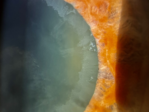
Cataract Surgery in Pseudoexfoliation Syndrome
All content on Eyewiki is protected by copyright law and the Terms of Service. This content may not be reproduced, copied, or put into any artificial intelligence program, including large language and generative AI models, without permission from the Academy.
Pseudoexfoliation (PXF) syndrome is characterized by the deposition of distinctive fibrillar material in the anterior segment of the eye. PXF is associated with the formation of dense nuclear cataracts as well as zonular compromise, and surgeries have a higher risk of developing complications.
PEX may be associated with potential serious ocular complications including rapidly progressive open angle glaucoma, corneal decompensation secondary to PEX keratopathy, spontaneous subluxation or luxation of the lens, cilio-lenticular angle closure glaucoma, and complications during cataract surgery.
Disease Entity
Cataract is the leading cause of blindness globally, particularly in developing countries. Currently, it accounts for 51% of 39 million blind people worldwide, 90% of them living in low- and middle-income countries. Cataract surgery in Pseudoexfoliation Syndrome have a higher risk of complications related to poor dilation and unstable zonules.
PXF syndrome is a multifactorial, genetically determined, age-related and environmentally influenced disorder of the elastic fiber structure, characterized by excessive production and accumulation of an elastotic material within a multitude of intra and extraocular tissues.
Disease
Pseudoexfoliative material is a grey-white fibrillary substance deriving from abnormal extracellular matrix metabolism in ocular and other tissues. The material is deposited on various ocular structures including the lens capsule , zonular fibres, iris, trabeculum and conjunctiva. Pseudoexfoliative material has been found in skin and visceral organs, leading to the concept that PXS is the ocular manifestation of a systemic disorder. PXS is associated with an increased prevalence of high-tone hearing loss and cardiovascular disorders.
PEX may be present unilaterally or bilaterally. PEX is strongly associated with raised intraocular pressures up to 44% of patients and subsequent development of pseudoexfoliation glaucoma, making it the most common identifiable cause of secondary open-angle glaucoma. PEX is also associated with technically challenging cataract surgery: PEX eyes dilate poorly and may have unstable lens zonules, which may lead to a higher risk of complications such as capsular bag rupture, zonular dialysis, and loss of vitreous.
Etiology
The pathogenesis of PXF Syndrome is multifactorial, but in some populations almost all patients with PXS have single nucleotide polymorphisms (SNPs) in theLOXL1 gene on chromosome 15, which codes for an enzyme that is involved in cross-linking of tropoelastin and collagen and is therefore important for the formation and maintenance of elastic fibres and extracellular matrix. However, these SNPs are common in the general population and most individuals with the SNPs do not develop PXS.
Risk Factors
- Old age (strongest risk factor; PEX rarely occurs below the age of 50)
- Race (nordic and Eastern Mediterranean populations have increased risk)
- Female sex (possible risk factor with more recent studies showing equal prevalence in males and females)
- Darker iris pigmentation (possible risk factor with conflicting results in studies)
- Low consumption of dietary anti-oxidants
PEX may be associated with potential serious ocular complications including rapidly progressive open angle glaucoma, corneal decompensation secondary to PEX keratopathy, spontaneous subluxation or luxation of the lens, cilio-lenticular angle closure glaucoma, and complications during cataract surgery.
General Pathology
Zonular weakness is a characteristic feature of PEX, and loosening of the zonular attachments to the basement membrane of the ciliary body is considered to be primarily responsible for this.
Preoperative Diagnosis
Diagnosis is made using slit lamp biomicroscopy and intraocular pressure measurement. Deposition of white fluffy material on the anterior lens capsule and pupillary margin and iris transillumination defects can be visualized in many cases. Gonioscopy may show increased pigment deposit on the trabecular meshwork.
Signs of reduced zonular integrity should be explored in order to assess the risk of complications during surgery. Typical signs of zonular weakness are phacodonesis, with eye movements, and sometimes a reduction or increase of the anterior chamber depth due to a anterior or posterior shift of the lens.
Poor pupil dilation is expected in most patients with PXF due to extracellular infiltration and degeneration of the dilator and sphincter muscles with atrophy of the pigment epithelium and stroma.
Variations of endothelial cell morphology and density are expected to be funded preoperatively in patients with PXF, and they may be responsible for an increased risk of corneal decompensation after surgery. Specular microscopy can be obtained before surgery.
Intraoperative Signs
In the era of phacoemulsification, PXF still represents a relevant challenge for the surgeon. The concurrent presence of poor pupil dilation and zonular weakness is responsible for the increased risk of intraoperative complications that may occur during surgery.
Management
Minimum sufficient pupil dilation (4–5 mm) may be achieved using a highly viscous cohesive ophthalmic visco- surgical device (OVD). Additional pupillary expansion devices may be used such as rings or hooks to achieve adequate pupillary dilation intraoperatively. Use of capsular support during surgery such as used of capsular hooks may be needed if zonular compromise is noted. Additionally, capsular tension rings and segments may be used to further stabilize the capsular bag prior to intraocular lens implantation.
- Large capsulorhexis: A capsulorhexis of 5.5 to 6.0 mm can simplify the extraction of nuclear pieces from the capsular bag and reduce potential for capsular phimosis in the future.
- Adequate hydrodissectio: An effective hydrodissection technique will help the nucleus to rotate freely and prevent further damage to the already weakened zonules. Though if severe zonular compromise is noted, rotation of the nucleus may need to be avoided until a quarter or more of the nucleus is removed in order to reduce further zonular stress.
- Irrigate the anterior chamber angle at the end of the surgery: Irrigating the angle can mobilize the PXF material and OVD from the trabecular meshwork and keep the IOP in check postoperatively


