Cherry-Red Spot
All content on Eyewiki is protected by copyright law and the Terms of Service. This content may not be reproduced, copied, or put into any artificial intelligence program, including large language and generative AI models, without permission from the Academy.
Cherry red spot is a significant fundoscopic finding in the macula, observed in central retinal artery occlusion (CRAO) and a variety of lipid storage disorders.
Disease Entity
Disease

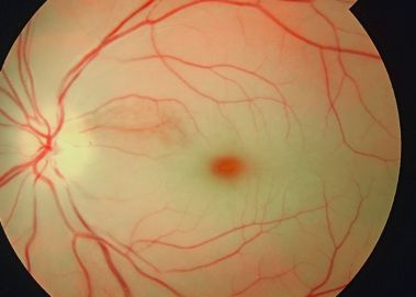
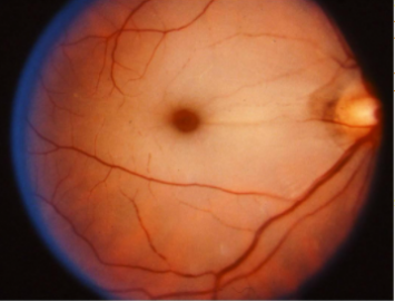
Cherry-red spot (CRS) at the macula is a clinically significant sign observed on fundus examination in a variety of pathological conditions[2], including retinal infarction, retinal ischemia. as well as a variety of lysosomal storage disorders.[3] The term refers to the appearance of a red-tinted region at the center of the macula surrounded by retinal opacification[3], and it consists a clinical sign usually present within the context of thickening and loss of transparency in the posterior pole of the retina.[4] The fovea retains relative transparency as it is the thinnest part of the retina and is devoid of ganglion cells. The fovea receives its blood supply from the choroid, which is supplied by the long and short posterior ciliary arteries.[5] The appearance of a cherry red spot can result from retinal edema most commonly due to central retinal artery occlusion or traumatic retinal ischemia, which cause the perimacular tissue of the retina to appear translucent, while the fovea maintains its normal color.[6][4] In the case of lipid storage diseases, the lipids are stored in the ganglion cell layer of the retina, giving the retina a white appearance. As ganglion cells are absent at the foveola, this area retains relative transparency and contrasts with the surrounding retina.[4] Cherry-red spot is an important fundoscopic sign, and when found with key clinical features and a good history, often guides one to the diagnosis of the disease.[2] It should be noted that as a condition resolves or evolves over the course of time, the appearance of a cherry-red spot diminishes and may not be present; therefore, absence of a cherry red spot does not indicate that a disease is not present.[3]
History
The term “cherry-red spot” was first introduced by American neurologist Bernard Sachs in 1887, when describing the fundus appearance of a child with "amaurotic familial idiocy" in his publication on “arrested development with special reference to its cortical pathology”. [6] The patient was confirmed upon neuropathologic examination to have lipid storage in the brain. [7] Bernard Sachs acknowledged that his patient had also been examined by ophthalmologist Herman Joseph Knapp, who practiced in both New York and Berlin, and described the striking retinal findings at an ophthalmology meeting in Heidelberg and used the term “cherry red color” to describe the fovea.[6] . Knapp had initially believed that the cherry red spot was a benign fundoscopic finding, but later on realized its grave implications. [6]
Etiology
The causes of cherry-red spot in the macula can be categorized into 6 groups, according to etiological factors:
Vascular
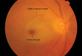
- Central retinal artery occlusion (CRAO) - This disease entity characteristically presents as a sudden onset of painless, unilateral visual loss in elderly patients, most commonly caused by an embolism, which blocks the central retinal artery.[3] The embolus may be composed of cholesterol, fibrin-platelet, or calcium and usually originates from the carotid plaque and less commonly from the heart or the aorta. Inflammatory conditions like giant cell arteritis (GCA), may also cause CRAO. Other causes of CRAO include myxoma or vegetations of the cardiac valves, thrombophilic disorders, and retinal migraine. Non-embolic vascular causes of retinal artery occlusion include nocturnal arterial hypotension, proatherogenic states such as hyperhomocystenemia,, factor V Leiden, protein C and S and anti-thrombin deficiencies, anti-phospholipid antibodies or prothrombin gene mutations, sickle cell disease, paraneoplastic syndromes and transient vasospasm.[8][9] Cherry-red spot has been found to be present on the initial examination in 90% of permanent CRAO cases.[3]
- Acute occlusion of the retinal circulation causing outer retinal whitening and a cherry-red spot at the macula following cataract surgery (phacoemulsification)[10]
- Intraorbital hemorrhage or mass may compromise the choroidal and retinal circulation and lead to a cherry-red spot at the macula
- Macular infarction due to various causes

Metabolic Storage Diseases
- Tay-Sachs disease (GM2 gangliosidosis type 1, infantile amaurotic familial idiocy, B variant GM2 gangliosidosis)- This is the most common type of gangliosidosis due to deficiency of the alpha subunit of hexosaminidase A on chromosome 15q23 and is associated with mutations in the gene HEXA. The disease has an onset of the symptoms in the infancy. There is an abnormal accumulation of gangliosides in the brain and ganglion cell layer of the retina. A cherry-red spot is present in approximately 90% of the patients affected with the disease.
- GM2 gangliosidosis type 2 or Sandhoff disease affecting gene HEXB
- Niemann-Pick disease - Type A, B, C, D disease
- GM1 gangliosidosis type 1 (Generalized gangliosidosis or Landing disease), in which a cherry-red spot in the macula occurs in around 50% cases. It is an autosomal recessive disorder caused by mutations in GLB1.
- Sialidosis or mucolipidosis type 1 is a lysosomal storage disease cause by alpha-neuraminidase deficiency (NEU1 gene mutation) and presents with ataxia, movement disorders, nystagmus, and myoclonic seizures. Intellect is usually normal, and somatic features are generally not seen. Two types have been described according to the age of onset and severity of the disease: Sialidosis type I or cherry-red spot myoclonus syndrome is characterized by onset in the 2nd or 3rd decade, gait disturbances, myoclonus, ataxia, tremor, a cherry-red spot at the macula, and seizures. The intellect and life expectancy are usually not affected, though the patient eventually needs wheelchair assistance with time. Almost all the patients have the cherry-red spot at the macula.
- Farber disease (Farber lipogranulomatosis) is a rare autosomal recessive (AR) disorder characterized by Ceramidase or acid ceramidase (chromosome 8p22) deficiency, affecting the GLA gene . The typical triad consists of painful swollen joints, subcutaneous nodules (lipogranulomas), and weak cry or hoarse voice due to laryngeal nodules. Other features include a developmental delay, visceromegaly, respiratory involvement, and neurological features, including quadriplegia, seizures, and myoclonus.
- Metachromatic leukodystrophy is another rare (AR) lysosomal storage disorder described by arylsulfatase A or deficiency, resulting in abnormal collection of sulfatides in myelin-producing cells that leads to progressive destruction of white matter (leukodystrophy). A cherry-red spot at the macula is occasionally a finding in fundoscopy along with progressive decline in intellectual and motor functions, peripheral neuropathy resulting in diminished sensations, hearing loss, incontinence, loss of speech and mobility, blindness, unresponsiveness, and eventual death.
- Galactosialidosis (Goldberg Cotlier syndrome or neuraminidase deficiency with beta-galactosidase deficiency or cathepsin A deficiency) is an AR disorder due to a mutation in the CTSA gene coding for cathepsin A. Ocular features include a cherry-red spot at the macula, corneal clouding, and conjunctival telangiectasia.
Inflammation
- Retinitis involving the central retina, including progressive outer retinal necrosis, subacute sclerosing panencephalitis. [3]
Drug Toxicity
- Quinine
- Carbon monoxide
- Dapsone poisoning associated with hemolytic anemia and peripheral neuropathy
- Methanol
- Intravitreal gentamicin
Trauma
- Commotio retinae - Acute retinal opacification following blunt trauma, also known as Berlin's edema, can cause a pseudo-cherry red spot, as a result of peripheral whitening of the retina that involves the macula.
- Optic nerve avulsion[12]
- Saturday night retinopathy, which refers to the occlusion of the retinal artery or the ophthalmic artery as a result of collapse of orbital vessels due to self-induced sustained direct orbit compression following headrest during prolonged prone body position.[13][14]
- Leukemic infiltrates of the optic nerve head may cause CRAO and cherry red spot
Risk Factors
Major risk factors of developing cherry-red spot at the macula due to CRAO include a variety of predisposing clinical conditions that can be divided into non-arteritic and arteritic.
- Nonarteritic : More than 90% of CRAOs are nonarteritic in origin, with Ipsilateral carotid artery atherosclerosis being the most common cause of retinal artery occlusion with a prevalence as high as 70% reported among patients with CRAO or branch retinal artery occlusion. Other non-arteritic causes of retinal artery occlusion are cardiogenic embolism, hematological conditions (sickle cell disease, hypercoagulable states, leukemia, lymphoma, etc.), and other vascular diseases, such as carotid artery dissection, Moyamoya disease, and Fabry disease.[15]
- Arteritic : CRAO of arteritic origin is mostly caused by giant cell arteritis, as well as other vasculitis disorders such as Susac syndrome, systemic lupus erythematosus, polyarteritis nodosa, and granulomatosis with polyangiitis.[15]
Pathophysiology
The macula is histologically characterized by an area consisting of more than one layer of ganglion cell layers, and the ganglion cell layer is thick at the macula. In disease entities causing loss of transparency (whitening or opacification) of the inner retina, the reddish color of the vascular choroid and pigmentation of retinal pigment epithelium (RPE) is not visible through the opacified retina. However, the foveola is devoid of the inner retinal layer. The retinal layers present at the foveola are (from vitreal to the sclera)- internal limiting membrane, outer nuclear layer, external limiting membrane, photoreceptor layer, and RPE. Inner retinal layers are absent in mature foveola. The vascular supply of the fovea is from the choroid and is not affected with occlusion of retinal vessels. Thus, the foveola, the thinnest part of the central retina, does not lose its transparency during inner retinal ischemia.
Thus, when there is inner retinal opacification, the reddish color of the vascular choroid and the RPE remain visible through the foveola, which is surrounded by an area of the white/opacified retina, giving rise to the typical cherry-red spot. The size of the cherry-red spot depends on the size of the foveola. There are several causes for opacification of the inner layers of the retina, including from deposition of compounds, as in storage diseases, or ischemia from vascular compromise involving the retina but not the underlying choroidal circulation.
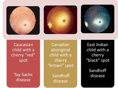
As the color of the choroid and RPE varies with racial variation, the color of the foveola may vary according to race. A true cherry-red spot presents in Caucasian individuals. Variations of the red-cherry spot are observed in non-Caucasian patients; the color of foveola was brown, a cherry-brown spot has been described in a Canadian aboriginal child with Sandhoff disease, due to the brown color of the fovea, whereas a cherry-black spot was reported in a patient of East Indian descent with Sandhoff disease., as a result of the black fovea color. Thus, an alternate term of 'perifoveal white patch' has been suggested. In cases of ophthalmic artery occlusion, there is a compromise to the vascular supply to retina, choroid, and optic nerve head circulation. There is retinal opacification without a cherry-red spot and severe vision loss with loss of light perception or accurate projection of rays.
Opacification of the inner retina can occur as a result of retinal ischemia due to occlusion of the central retinal artery . The retinal whitening is typically most prominent in the posterior pole, and the opaque retina obscures the details of the choroidal vessels. The whitening of the retina becomes less prominent a little peripheral to the arcades, which may be related to the reduced inner retinal thickness at the peripheral retina. There is a cessation of axoplasmic transport and then the opacification of both the ganglion cell and the nerve fiber layer in a CRAO.
A variety of lipid storage disorders can produce deposition / accumulation of material at the level of the ganglion cell layer can other lipid storage disorders (sphingolipidoses), leading to progressive abnormal excessive intracellular accumulation of various glycolipids/phospholipids in the ganglion cell layer, that cause opacification of the inner retina. The foveola is devoid of the ganglion cell layer and hence remains transparent and allows transmission of the color of the choroid and the RPE. However, with the increasing age, the damaged ganglion cells are lost, and the cherry-red spot becomes less prominent. In such cases, there is consecutive optic atrophy and pallor of the optic disc due to the degeneration of the ganglion cell layer and nerve fiber layer.
Primary prevention
Prevention for retinal ischemia can include measures to reduce cardio or cerebrovascular diseases.
Diagnosis
Ruling out serious life-threatening or sight-threatening conditions is of paramount importance in a patient with a cherry-red spot at the macula, thus requiring a thorough evaluation and management of such patients.[3] Acute loss of vision with a cherry red spot should raises concern of risk for a stroke occurring. Depending on the institution, initiating consultation with a stroke team or neurology is important. Also important is assessing blood pressure.
History
- Preceding history of amaurosis fugax (transient vision loss lasting seconds to minutes, but that may last up to 2 hours)
- Medical history of cardiovascular disease, thromboembolic disorders, cerebrovascular disease, proatherogenic states such as hyperhomocystenemia, factor V Leiden, protein C and S and anti-thrombin deficiencies, anti-phospholipid antibodies or prothrombin gene mutations, sickle cell disease, neoplastic disease
- Smoking
- Family history of lipid/lysosomal storage disorders or metabolic disease
- Ocular trauma
- Face-down surgical procedures
- Drug use
Signs

Fundoscopic features that present along with cherry-red spot at the macula include:
- Yellow-white opacified appearance of the retina in the posterior pole, due to ischemic necrosis affecting the inner half of the retina, which vanishes in 4-6 weeks.
- Cloudy swelling
- Attenuated retinal arteries
- Thinning, dilation or normal appearance of retinal veins
- Segmentation or "box-carring” (“cattle trucking”) of the blood column due to arteriolar narrowing in severe obstruction.[9]
- Pallor of the optic disc (39% in CRAO cases) in later stages, as well as visible absence of nerve fiber layer in the region of optic disc[17]
- Presence of emboli (20-40% in CRAO cases)
- Pigmentary changes indicate that choroidal circulation is also involved.[17]
- Presence of a Hollenhorst plaque, which is a clinical finding in cases of central retinal artery occlusion, or one of its branches (BRAO) caused by a lodging embolus deriving from a cholesterol plaque. [8]
Symptoms
- Sudden onset painless vision loss occurring over several seconds
- Symptoms of giant cell arteritis in older patients including headache, jaw claudication, scalp tenderness, proximal muscle and joint aches, anorexia, weight loss, fever
- Transient migraine due to vasospasm
- Neurological symptomatology indicative of lysosomal storage disorders, metabolic disease.
Clinical Diagnosis
- Spectral-domain Optical Coherent Tomography (OCT) has been proposed as one modality that might be used to diagnose and monitor CRAO. In CRAO, there is an observed increase in intensity of inner retinal layers compared with age-matched controls, and it corresponds to the layers supplied by central retinal arteries.[18] OCT can be correlated with visual prognosis.[18] Incomplete CRAO shows minimal retinal architectural disruption and inner layer hyper-reflectivity without retinal edema. Subtotal CRAO demonstrated inner macular thickening and loss of organization of the inner retina, and total CRAO demonstrated marked inner retinal thickening and subfoveal choroidal thinning. [19][20] In the chronic phase, there is a corresponding thinning of the inner retinal layers.
- Fluorescein Angiography may be a prognostic test in CRAO, as poor perfusion on fluorescein angiography has been associated with lower vision than exudative and mixed perfusion. However, this finding does not influence therapy. [21]Normal choroidal filling begins 1-2 seconds before retinal filling and is complete within 5 seconds of dye appearance in healthy eyes. [21]A delay of 5 or more seconds is seen in 10% of patients.[21] Ophthalmic artery occlusion or carotid artery obstruction should be considered if choroidal filling is significantly delayed. A delay is noted in arteriovenous transit time (reference range, < 11 seconds), as well as a delay in retinal arterial filling. Arterial narrowing with normal fluorescein transit is observed after recanalization.[21]
- Optical Coherent Tomography Angiography provides structural and functional (blood flow) information at a fixed point, but is not useful to appreciate leakage from vessels. [29] OCTA shows decreased vascular perfusion in superficial and deep retinal plexus that corresponds to poor perfusion on fluorescein angiography.[22] However, unlike fundus fluorescein angiography, OCTA cannot demonstrate a delay in transit time. [23]
- Fundus autofluorescence : in the acute phase pf CRAO, fundus autofluorescence in ischemic areas is decreased because of retinal edema blocking the normal RPE. Eventually, this could return to normal baseline or may be associated with increased autofluorescence owing to a window defect created by the thinned-out inner retinal layers. [24]
- Electroretinography shows a diminished b-wave corresponding to Muller and/or bipolar cell ischemia.
Diagnostic procedures
- Blood pressure and pulse monitoring, particularly to detect atrial fibrillation, possibly with Holter monitor
- Carotid Doppler Ultrasonography may be used to evaluate for atherosclerotic plaque; flow
- ECG to detect possible atrial fibrillation , arrhythmia or other cardiac disease.
- Echocardiography is usually performed when there is a specific indication such as a history of rheumatoid fever, known cardiac valvular disease.
- Chest X-ray to investigate for sarcoidosis, tuberculosis, left ventricular hypertrophy in hypertension
- Magnetic resonance imaging, as approximately 20% of patients with a CRAO also have cerebral ischemia; therefore a brain MRI may reveal concurrent cerebral ischemia in patients without accompanying neurological symptoms.[25]
- Magnetic resonance angiography (MRA) of the head and neck may be more accurate in detecting vascular occlusive disease.
- Computerized tomography (CT) or computerized tomography angiography (CT/CTA) or MRI/MRA of the neck may be needed for carotid dissection
Laboratory test
- Complete blood count CBC (anemia, leukemia, polycythemia, platelet disorders)
- urea and electrolytes
- Erythrocyte sedimentation rate (ESR) to detect the remote possibility of giant cell arteritis in elderly patients
- C-reactive protein
- Hypercoagulable state evaluation (eg, factor V Leiden, prothrombin mutation, homocysteine levels, fibrinogen, antiphospholipid antibodies, prothrombin time/activated partial thromboplastin time [PT/aPTT], serum protein electrophoresis, among others)
- Fasting blood sugar, cholesterol, triglycerides, and lipid panel to evaluate for atherosclerotic disease
- Homocysteine levels
- Plasma protein electrophoresis (sickle-cell anemia)
- Blood cultures to evaluate for suspected bacterial endocarditis and septic emboli
- Antibodies
- Genetic testing, molecular analysis of cells or tissues, and enzyme assays to investigate possible lipid-storage disorders in younger patients
Differential diagnosis
A macular hemorrhage or hole, with resulting enhanced red contrast relative to the surrounding retina, may be confused with a cherry red spot.[6]
Management
General treatment
Cherry-red spot at the macula is a clinically significant fundoscopic finding that usually denotes an ocular emergency especially when observed in a patient who presents with sudden, painless, vision loss of one eye . The general management of a cherry-red spot sign is determined by the underlying pathological condition that causes the appearance of the fundoscopic finding.
In the acute setting of CRAO, treatment options are directed at resolving the CRAO and circulation, and thus maximizing visual outcome. Experimental studies suggest no detectable retinal damage in primate models with CRAO if retinal blood flow is restored within 90 minutes. Subsequent partial recovery may be possible if ischemia is reversed within 240 minutes. However, occlusions lasting longer than 240 minutes produce irreversible damage. The treatment approach is attempted within the first 24 hours.
For various storage diseases, the management requires a team of various specialties. Generalized pearls for the management of such patients include avoiding/reducing intake of specific molecules that lead to accumulation within ganglion cells, enzyme replacement therapy, and symptomatic management.
Medical therapy
IOP reducing pharmacological agents
- IV acetazolamide
- IV mannitol
- Topical antiglaucoma medications
Vasodilation agents
- Pentoxifylline
- Inhalation of carbogen
- Sublingual isosorbide dinitrate
Thrombolytic therapy to dissolve clot
- IV or intra-arterial recombinant tissue plasminogen activator (rt-PA) is delivered through the catheterization of the ophthalmic artery or supraorbital artery and can reach the CRA in doses 100 times greater than by systemic administration. Vision improvement is noted in 50% of the patients.[15]
IV methylprednisolone
(only in cases of arteritic CRAO)
Hyperbaric oxygen therapy
Procedures / Surgery
- Ocular massage : it involves in and out movement with the use of a three-mirror (Goldmann) contact lens or digital massage, at an attempt to mechanically dislodge the obstructing embolus. Increase pressure should be applied for 10-15 seconds followed by sudden release. Ocular massage can cause arterial dilatation andi mprove retinal perfusion by an 86% increase in flow volume.[15]
- Anterior chamber paracentesis : performing this procedure can provide sudden decrease in intraocular pressure, and aids the perfusion pressure behind the obstruction to dislocate the obstructing embolus.[15]
- Transluminal Nd:YAG laser embolysis : the procedure has been advocated for CRAO or BRAO, in cases when the occluding embolus is visible an be performed to lyse or dislodge Shots of 0.5–1.0 mJ or higher are applied directly to the embolus using a fundus contact lens, in order to lyse or dislodge the occluding embolus. [15]Embolectomy has been said to occur if the embolus is ejected into the vitreous via a hole in the arteriole. The main complication is vitreous haemorrhage.
- Pars plana vitrectomy : Surgical removal of the clot
Complications
Neovascularization is a complication that may occur in patients with CRAO with an incidence that varies from 3.0-18.8%. It may involve the retina, iris, or iridocorneal angle. Moreover, the detection of neovascularization following CRAO has ranged from as early as the day of presentation to 2 years after the CRAO diagnosis. The association of CRAO and the development of neovascular glaucoma is thought to occur in about 5%; however, a prospective study established a temporal relationship between CRAO and neovascular glaucoma in 15% of cases. These results are corroborated by a retrospective study that showed a mean of 8.5 weeks from diagnosis to clinically evident neovascularization.9 However, a prospective study of 232 eyes found neovascularization in 2.5% of cases, and the authors found that there was no causal relationship.[26]
With frequent visits, many cases of neovascularization can be managed early with treatments such as panretinal photocoagulation to decrease retinal oxygen demand and off-label intravitreal bevacizumab. Since neovascularization can occur early, regular follow-up appointments should be required, especially within the first 4 months.[26]
Prognosis
The initial management approach described above may provide some benefit; however, there is no clear evidence at this point that these measures result in improvement of the patient's final visual acuity and have the best chance when performed shortly after the diagnosis of CRAO. Vision loss is permanent due to infarct of inner retina, as irreversible damage to sensory retina develops only after 90-100 minutes of complete CRAO. Vision is 20/400 or worse in 66% of cases.
References
- ↑ https://commons.wikimedia.org/wiki/File:Cherry_red_spot_in_patient_with_central_retinal_artery_occlusion_(CRAO).jpg
- ↑ Jump up to: 2.0 2.1 Suvarna J C, Hajela S A. Cherry-red spot. J Postgrad Med 2008;54:54-7
- ↑ Jump up to: 3.0 3.1 3.2 3.3 3.4 3.5 3.6 Tripathy K, Patel BC. Cherry Red Spot. [Updated 2021 Feb 14]. In: StatPearls [Internet]. Treasure Island (FL): StatPearls Publishing; 2021 Jan-. Available from: https://www.ncbi.nlm.nih.gov/books/NBK539841/
- ↑ Jump up to: 4.0 4.1 4.2 General Practice Notebook
- ↑ USMLE First AID 2010 page 417
- ↑ Jump up to: 6.0 6.1 6.2 6.3 6.4 Leavitt, J. A., & Kotagal, S. (2007). The “Cherry Red” Spot. Pediatric Neurology, 37(1), 74-75 doi:10.1016/j.pediatrneurol.2007.04.011
- ↑ Leavitt, J. A., & Kotagal, S. (2007). The “Cherry Red” Spot. Pediatric Neurology, 37(1), 74–75. doi:10.1016/j.pediatrneurol.2007.04.011
- ↑ Jump up to: 8.0 8.1 Kaufman EJ, Mahabadi N, Patel BC. Hollenhorst Plaque. [Updated 2021 Feb 25]. In: StatPearls [Internet]. Treasure Island (FL): StatPearls Publishing; 2021 Jan-. Available from: https://www.ncbi.nlm.nih.gov/books/NBK470445/
- ↑ Jump up to: 9.0 9.1 Varma DD, Cugati S, Lee AW, Chen CS. A review of central retinal artery occlusion: clinical presentation and management. Eye (Lond). 2013 Jun;27(6):688-97. doi: 10.1038/eye.2013.25. Epub 2013 Mar 8. PMID: 23470793; PMCID: PMC3682348.
- ↑ Central retinal artery occlusion after phacoemulsification under peribulbar anaesthesia: S Rodríguez Villa R Salazar Méndez M Cubillas Martín M Cuesta García Eur J Ophthalmol. 2021 Mar;31(2):NP77 PMID: 26652970 DOI: 10.1016/j.oftal.2015.10.003
- ↑ Learning About Tay-Sachs Disease [Internet]. 2011 [cited 2013 Oct 20] Available from: http://www.genome.gov/10001220
- ↑ Sudden occlusion of the retinal and posterior choroidal circulations in a youth. Brown GC, Magargal LE.Brown GC, et al. Am J Ophthalmol. 1979 Oct;88(4):690-3. doi: 10.1016/0002-9394(79)90666-4.
- ↑ Self-induced Orbital Compression Injury: Saturday Night Retinopathy. Williams AL, Greven M, Houston SK, Mehta S.Williams AL, et al. JAMA Ophthalmol. 2015 Aug;133(8):963-5. doi: 10.1001/jamaophthalmol.2015.1114.
- ↑ Williams AL, Greven M, Houston SK, Mehta S. Self-induced Orbital Compression Injury: Saturday Night Retinopathy. JAMA Ophthalmol. 2015;133(8):963–965. doi:10.1001/jamaophthalmol.2015.1114
- ↑ Jump up to: 15.0 15.1 15.2 15.3 15.4 15.5 Sim, S., & Ting, D. (2017, August). Diagnosis and Management of Central Retinal Artery Occlusion. American Academy of Ophthalmology. https://www.aao.org/eyenet/article/diagnosis-and-management-of-crao.
- ↑ Varma DD, Cugati S, Lee AW, Chen CS. A review of central retinal artery occlusion: clinical presentation and management. Eye (Lond). 2013 Jun;27(6):688-97. doi: 10.1038/eye.2013.25. Epub 2013 Mar 8. PMID: 23470793; PMCID: PMC3682348
- ↑ Jump up to: 17.0 17.1 Hayreh SS, Zimmerman MB. Fundus changes in central retinal artery occlusion. Retina. 2007 Mar;27(3):276-89. doi: 10.1097/01.iae.0000238095.97104.9b. PMID: 17460582.
- ↑ Jump up to: 18.0 18.1 Chen H, Chen X, Qiu Z, Xiang D, Chen W, Shi F, et al. Quantitative analysis of retinal layers' optical intensities on 3D optical coherence tomography for central retinal artery occlusion. Sci Rep. 2015 Mar 18. 5:9269.
- ↑ Chen H, Chen X, Qiu Z, Xiang D, Chen W, Shi F, et al. Quantitative analysis of retinal layers' optical intensities on 3D optical coherence tomography for central retinal artery occlusion. Sci Rep. 2015 Mar 18. 5:9269.
- ↑ Mehta N, Marco RD, Goldhardt R, Modi Y. Central Retinal Artery Occlusion: Acute Management and Treatment. Curr Ophthalmol Rep. 2017 Jun. 5 (2):149-159.
- ↑ Jump up to: 21.0 21.1 21.2 21.3 Gong H, Song Q, Wang L. Manifestations of central retinal artery occlusion revealed by fundus fluorescein angiography are associated with the degree of visual loss. Exp Ther Med. 2016 Jun. 11 (6):2420-2424.
- ↑ de Carlo TE, Romano A, Waheed NK, Duker JS. A review of optical coherence tomography angiography (OCTA). Int J Retina Vitreous. 2015. 1:5.
- ↑ Bonini Filho MA, Adhi M, de Carlo TE, Ferrara D, Baumal CR, Witkin AJ, et al. OPTICAL COHERENCE TOMOGRAPHY ANGIOGRAPHY IN RETINAL ARTERY OCCLUSION. Retina. 2015 Nov. 35 (11):2339-46.
- ↑ Mathew R, Papavasileiou E, Sivaprasad S. Autofluorescence and high-definition optical coherence tomography of retinal artery occlusions. Clin Ophthalmol. 2010 Oct 21. 4:1159-63.
- ↑ Dattilo M, Biousse V, Newman NJ. Update on the Management of Central Retinal Artery Occlusion. Neurol Clin. 2017 Feb. 35 (1):83-100.
- ↑ Jump up to: 26.0 26.1 Jung YH, Ahn SJ, Hong JH, et al. Incidence and Clinical Features of Neovascularization of the Iris following Acute Central Retinal Artery Occlusion. Korean J Ophthalmol. 2016;30(5):352-359. doi:10.3341/kjo.2016.30.5.352


