Wide Field Retinal Imaging Systems: Difference between revisions
No edit summary |
No edit summary |
||
| Line 4: | Line 4: | ||
|Category=Retina/Vitreous | |Category=Retina/Vitreous | ||
|Assigned editor=Neelakshi.E.Bhagatne | |Assigned editor=Neelakshi.E.Bhagatne | ||
|Reviewer= | |Reviewer=Neelakshi.E.Bhagatne | ||
|Date reviewed= | |Date reviewed=May 22, 2019 | ||
}} | }} | ||
= Introduction = | = Introduction = | ||
| Line 16: | Line 16: | ||
= The Evolution in Retinal Imaging = | = The Evolution in Retinal Imaging = | ||
Retinal imaging techniques have evolved at a remarkable pace in the last two decades. Wide field imaging (WFI) and ultra wide field imaging (UWFI) are now increasingly popular. WFI refers to imaging beyond 50 degrees field area. UWFI systems can image upto 200 degrees. They are well capable of imaging over 80% of the retinal surface area. Besides imaging, they also provide valuable information about the peripheral vasculature and other retinal lesions. | Retinal imaging techniques have evolved at a remarkable pace in the last two decades. Wide field imaging (WFI) and ultra wide field imaging (UWFI) are now increasingly popular. WFI refers to imaging beyond 50 degrees field area. UWFI systems can image upto 200 degrees. They are well capable of imaging over 80% of the retinal surface area. Besides imaging, they also provide valuable information about the peripheral vasculature and other retinal lesions that might otherwise be missed with traditional imaging systems. | ||
The modern systems allowing 100° to 200° view of fundus include: | The modern systems allowing 100° to 200° view of fundus include: | ||
| Line 33: | Line 33: | ||
* Possibility of image transmission via electronic route | * Possibility of image transmission via electronic route | ||
* Better acquisition in eyes with cataract than a traditional fundus camera | * Better acquisition in eyes with cataract than a traditional fundus camera | ||
* Simultaneous imaging of central and peripheral retina | * Simultaneous imaging of central and peripheral retina; use of UWF FA to evaluate peripheral retinal ischemia in patients with diabetic macular edema and other vascular complications in the central macula | ||
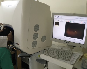
[[File:Optos.JPG|thumb|Optos 200x camera]] | [[File:Optos.JPG|thumb|Optos 200x camera]] | ||
Revision as of 10:32, May 22, 2019
All content on Eyewiki is protected by copyright law and the Terms of Service. This content may not be reproduced, copied, or put into any artificial intelligence program, including large language and generative AI models, without permission from the Academy.
Introduction
Retinal imaging involves creating a two-dimensional image of the three-dimensional (3D) retinal tissue. Such retinal imaging techniques are indispensable for diagnosis and management of disease processes in ophthalmic practice. These help ophthalmic practioners directly to view the retinal disease and plan treatment according to the pathology. Retinal imaging techniques are useful in diagnosis and management of ocular and systemic disorders like diabetic retinopathy, hypertensive retinopathy, age-related macular degeneration, vascular pathologies (vascular occlusions, vasculitis, etc), retinal detachments, glaucomas, systemic infections, leukemias, systemic malignancies with ocular metastasis etc.
The Past in Retinal Imaging
Dating back to the early 20th century, ophthalmologists relied upon the tedious process of capturing fundus images and fundus fluorescein angiography (FFA) on film rolls. The late 20th century and earlier part of 21st century saw advancements in retinal imaging techniques. Since the advent of digital imaging, all traditional fundus cameras have been replaced by digital systems.
The most impactful advancement in the recent years in retinal imaging has been the introduction of Optical Coherence Tomography (OCT). Since its introduction in the 1990s, much has changed in our understanding of many retinochoroidal diseases, in our approach to management of common conditions and in our definitions of treatment success and end-point in a lot of those diseases.1 OCT has been pivotal in identifying and understanding may diseases like Idiopathic macular hole and vitreomacular traction syndrome, myopic traction maculopathy, macular telangiectasia type 2 and pachychoroid spectrum of diseases.In many instances, OCT has obviated the need for repeated fluorescein angiography for continued management decisions. High speed OCT machines make it possible to acquire large number of A-scans and to convert them into highly resolved volume data. High speed OCT machines and some novel algorithms of OCT signal analysis has also led to the development of OCT angiography (OCT-A).
The Evolution in Retinal Imaging
Retinal imaging techniques have evolved at a remarkable pace in the last two decades. Wide field imaging (WFI) and ultra wide field imaging (UWFI) are now increasingly popular. WFI refers to imaging beyond 50 degrees field area. UWFI systems can image upto 200 degrees. They are well capable of imaging over 80% of the retinal surface area. Besides imaging, they also provide valuable information about the peripheral vasculature and other retinal lesions that might otherwise be missed with traditional imaging systems.
The modern systems allowing 100° to 200° view of fundus include:
- Pomerantzeff camera
- Retcam (Clarity Medical Systems, Inc., Pleasanton, CA, USA)
- Panoret-1000™ camera (Medibell Medical Vision Technologies, Haifa, Israel),
- Optos® camera (Optos PLC, Dunfermline, UK)
- Heidelberg Spectralis with the Staurenghi lens (Ocular Staurenghi 230 SLO Retina Lens; Ocular Instruments Inc, Bellevue, WA, USA)
Today, retinal imaging includes near histopathological examination of retina, viewing much beyond the equator, non-invasive ways of visualizing the retinal vasculature, health of (RPE) Retinal Pigment Epithelium, photoreceptor density, quantifying various fundus pigments, the blood flow, oxygenation, the molecular transport and an ever increasing number of highly exciting things.1
Advantages of modern digital WFI and UWFI systems:2
- Enhanced resolution
- Shorter image processing time
- Faster image acquisition
- Ease of image duplication, manipulation and
- Possibility of image transmission via electronic route
- Better acquisition in eyes with cataract than a traditional fundus camera
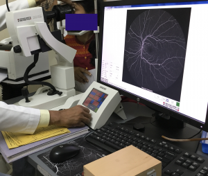
- Simultaneous imaging of central and peripheral retina; use of UWF FA to evaluate peripheral retinal ischemia in patients with diabetic macular edema and other vascular complications in the central macula
Confocal scanning laser ophthalmoscopy imaging (CSLO) systems
Advances in high-resolution OCT currently under development promise an even more detailed fundus representation. The integration of the confocal scanning laser ophthalmoscope (CSLO) and OCT has produced a dynamic new instrument, the OCT ophthalmoscope, which simultaneously images the fundus in numerous ways with point to point correlation.2 CSLO systems use laser light to illuminate the retina, instead of bright flashes of light. This reduces scatter of light in images acquired.
Examples of cSLO-based ultrawidefield imaging (UWFI) systems include:
- the Optos® camera (Optos PLC, Dunfermline, UK),
- the Spectralis® (Heidelberg Retina Angiograph (HRA 2), Heidelberg Engineering, Germany).
Multimodal Imaging with WFI and UFWI systems
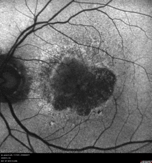
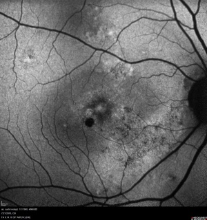
A great advantage offered by many of the present WFI and UWFI systems is the possibility of simultaneous acquisition of fundus fluorescein agniography (FFA), indocyanine angiography (ICGA), red-free photography, fundus photography, color fundus stereo imaging, adaptics optics CSLO, hyperspectral retinal imaging, fundus autofluorescence (FAF); including blue-reflectance (BAF), infrared reflectance (IRAF) or green reflectance (GAF).
| Available Platforms/Devices | Type of lens system | Principle | Field of view | Facilities available | |
|---|---|---|---|---|---|
| WFI | Heidelberg Spectralis | Non-contact | SD-OCT with CSLO | 55° (upto 105° with HRA 2) | FFA, ICGA, FAF (BAF and IRAF) |
| Contact | SD-COT with CSLO using Staurenghi Lens | 150° | FFA, ICGA, FAF (BAF and IRAF) | ||
| RetCam 3 | Contact | Optical light source to obtain high resolution | Field depends on lens used among the five changeable lens systems3:
-130° ( pediatric retina and adult anterior chamber), -120°( pediatric and young adult), -80°(high contrast pediatric and adult), -30° (high magnification) and Portrait (external imaging) |
FFA, ICGA | |
| UWFI | Optos Optomap | Noncontact | CSLO-based | 200 | FFA, FAF (GAF, IRAF) |
Applications of WFI and UWFI in Ocular Conditions
- Retinal Vascular occlusions
- Retinal Vasculitis
- Pediatric retinal conditions
- Posterior Uveitis - infectious and non-infectious
- Peripheral retinal detachments
- Peripheral polypoidal choroidal vasculopathy (Peripheral PCV)
- Adult onset foveomacular vitelliform dystrophy (AOFVD)
- Familial Exudative vitreoretinopathy (FEVR)
- Eales' disease
- Retinal detachments - exudative and tractional
- Screening for diabetic retinopathy and glaucoma
Limitations of WFI and UWFI
- Difficulty to precisely measure the retinal surface area in order to estimation size and dimensions of retinal lesions.
- Image artifacts
- Conversion of a 3D surface to 2D image is still a challenge in retinal imaging
The Future Direction
Advanced imaging techniques have revolutionized the fundus examination in ophthalmology. Future in retinal imaging is expected to be as exciting as the recent journey has been, if not more. Research prototype swept source OCT with speeds as high as 6700000 A scans/sec have been reported in recent literature. What this can do to the quality, speed and the quantity of data that can be acquired is something everyone is waiting to witness.With such high speed OCTs, 4-D intraoperative OCT may not be very distant. Recently, a frequency-swept light source called as the vertical cavity surface emitting laser was introduced with a very high imaging range of up to 50mm. It has been reported to image the entire eye, including anterior segment, lens, vitreous, retina, choroid and sclera in a single OCT – A 3D OCT image of the entire eye!
Ultra wide field indo cyanine green angiography is being looked forward to and is expected to provide more insights into diseases like CSC and polypoidal choroidal vasculopthy. Quantitative (AF) Auto Fluorescence is a way of quantifying the amount of AF emitted by the RPE. It has been shown that even normal looking and normal testing fundus in conditions like Stargardt disease show massive accumulation of autofluorescent material providing us new insight into disease and a possible means of prognosticating the patient. Adaptive Optics-Scanning Laser Ophthalmoscopes with fluorescein angiography filters are expected which will provide histopathological resolution of macular vascular architecture and may change the way we manage macular ischemia.
Conclusion
As advancements continue to develop in the field of retinal imaging, our understanding of ocular disease processes continues to improve. Newer technologies, which address the need of ophthalmologists towards achieving the proper diagnosis and appropriate management of disease entitities, have helped improve the patient care and management ultimately in our current ophthalmic practice.
References
- Atul Kumar. Retinal Imaging-Present and future. Off Sci J Delhi Ophthalmol Soc. 2017;27(4):241-242. doi:10.7869/djo.257.
- Yannuzzi LA, Ober MD, Slakter JS, Spaide RF, Fisher YL, Flower RW, Rosen R. Ophthalmic fundus imaging: today and beyond. AmJ Ophthalmol. 2004;137(3):511-524. doi:10.1016/j.ajo.2003.12.035.
- http://www.claritymsi.com/us/downloads/Lenses&Images_2009_ f.pdf as accessed on 4 May 2015.


