All content on Eyewiki is protected by copyright law and the Terms of Service. This content may not be reproduced, copied, or put into any artificial intelligence program, including large language and generative AI models, without permission from the Academy.
It is important for the oculoplastic surgeon, or anyone performing orbital surgery, to have a strong understanding vascular anatomy. While significant variability exists[1], this article will detail the standard vascular anatomy of the human orbit.
Arterial Supply to the Orbit
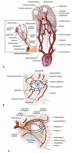
Blood supply to the orbital arises primarily from the ophthalmic artery - the first branch off of the internal carotid as it emerges from the cavernous sinus on the medial side of the clinoid process. The ophthalmic artery has many branches which may be separated into 2 groups: *Orbital Group *Ocular Group
Orbital Group
- Lacrimal artery: Runs along the lateral wall of the orbit and supplies the lacrimal gland. Terminal branches of the lacrimal artery include the superior and inferior lateral palpebral arteries which supply the lateral upper and lower eyelids and conjunctiva.
- Supraorbital artery: In the orbit supplies the supeior rectus and levator palpebral muscles. It then passes through the supraorbital foramen and its terminal branches supply the eyebrow and forhead.
- Anterior ethmoidal artery: In the orbit supplies the superior oblique muscle. Also supplies the anterior and middle ethmoidal cells, frontal sinus, lateral wall nose, and nasal septum.
- Posterior ethmoidal artery: Passes through the posterior ethmoidal canal, supplying the posterior ethmoidal cells.
- Internal palpebral artery: Terminal branches include superior and inferior medial palpebral arteries. These vessels supply the lacrimal sac and eyelids creating an anastomosis with the two lateral palpebral branches from the lacrimal artery.
- Frontal artery: Leaves the orbit at its medial angle above the trochlea and supplies the forehead and scalp creating an anastomosis with the supraorbital artery terminal branches.
- Nasal artery: Supplies the superior lacrimal sac and nose.
Ocular Group
- Short ciliary artery: Supply the choroid and cilliary processes.
- Long ciliary artery: Supply the anterior segment and form anastomoses/collateralization with extraocular muscles branches.
- Central retinal artery: Branches off of the posterior 1/3 of the ophthalmmic artery and enters the dural sheath of the optic nerve about 13mm behind the globe. Supplies the retina.
- Muscular arteries: Splits into superior and inferior branches. The superior branch supplies the levator palpebral muscle, superior rectus, superior oblique and a portion of the lateral rectus. The inferior branch supplies the medial rectus, inferior rectus and inferior oblique. This inferior branch also gives off most of the anterior ciliary arteries.
Note: Anastomoses to the periorbit are also formed via the facial and superficial temporal branches of the external carotid.
Orbital Veins
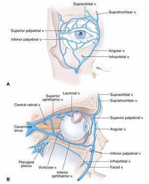
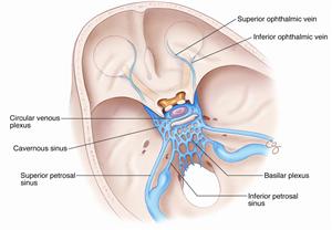
- Superior Ophthalmic Vein: Provides the main venous drainage of the orbit. Originates in the superonasal quadrant of the orbit and extends posteriorly through the medial part of the superior orbital fissure into the cavernous sinus.
- Inferior Ophthalmic Vein: Originates at the floor and medial wall of the orbit and provides drainage for the inferio orbit.
- Vortex Veins: Venous drainage of the uveal tract and pierce the sclera posterior to the equator of the globe. Superior vortex veins (lateral and medial) drain into the superior ophthalmic vein and the inferior vortex veins (lateral and medial) drain into the inferior ophthalmic vein.
References
- ↑ Hayreh SS. Orbital vascular anatomy. Eye (2006) 20, 1130–1144. doi:10.1038/sj.eye.6702377


