All content on Eyewiki is protected by copyright law and the Terms of Service. This content may not be reproduced, copied, or put into any artificial intelligence program, including large language and generative AI models, without permission from the Academy.
Surgical procedure
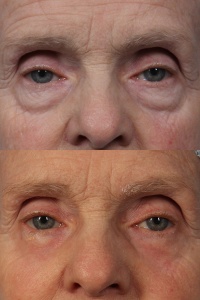
There are many varieties of lower blepharoplasty surgery. The earliest forms involved making an incision with a scalpel under the lash line of the lower eyelids and removing skin and or fat. More modern techniques often involve an incision behind the eyelid (transconjunctival approach) and combine a variety of instruments including monopoloar cautery and CO2 laser. Lower blepharoplasty techniques may including fat repositioning, fat grafting, and/or surgery to address the underlying orbicularis oculi muscle and/or Sub - Orbicularis Oculi Fat pads (SOOF). Additional details will be given below.
Background
The term “lower blepharoplasty” includes a collection of surgical techniques that aims to improve the appearance of the lower eyelids. Historically, lower blepharoplasty was a reductive procedure in which skin and/or fat was removed in order to reduce lower eyelid wrinkles, skin redundancy, and fat bulges. While fat and skin excision is still performed with modern lower blepharoplasty , present trends follow a tissue-preserving philosophy that may include orbital and sub-orbicularis fat repositioning and fat transfer techniques to restore apparent volume loss associated with facial aging. In the early 2000’s, hyaluronic acid-based dermal fillers emerged as an off-label means of lower eyelid and infra-orbital volumization. Laser energy and light-based treatments have also been applied to the lower eyelids, providing non-surgical lower blepharoplasty options or non-surgical adjuncts to incisional blepharoplasty.
Patient Selection
History
A thorough medical and ophthalmic history is obtained prior to cosmetic lower blepharoplasty including:
- Current illnesses
- Medication list (including anticoagulants, vitamins, herbal pills)
- Ophthalmic medications, lubricants, and contact lens wear
- Drug or latex allergies
- Dry eye symptoms
- Social history (smoking, occupation, sun exposure)
- Description of previous facial, ophthalmic, and eyelid surgery and procedures
- The patient’s goals and expectations are discussed
Physical Examination
- Visual acuity, pupillary exam, extraocular motility.
- Tear film and ocular surface evaluation
- Presence of Bell’s phenomenon
- Blink rate and strength
- Presence of lower eyelid laxity and canthal tendon dehiscence
- Presence of lagophthalmos
- Skin evaluation (Fitzpatrick skin type, rhytidosis, dyschromia, skin redundancy, lesions)
- Presence of steatoblepharon, infraorbital hollowing, tear trough deformity, malar fat atrophy
- Globe prominence and globe/maxilla relationship (presence of negative vector)
- Asymmetries, orbital dystopia
Standard view external photographs are obtained prior to surgery.
Indications
- Rhytidosis and lower eyelid dermatochalasis
- Relative steatoblepharon
- Pronounced nasojugal groove
- Infraorbital/malar deflation
- Malar mounds or festoons
- Lower eyelid asymmetry
Contraindications
- Unachievable patient goals / unrealistic expectations
- Coexisting severe or unstable medical conditions
- Active thyroid ophthalmopathy (relative contraindication)
- Uncontrolled dry eye syndrome
Surgical technique
An effective surgical rejuvenation of the lower eyelids addresses the patient’s concerns that correspond with anatomic issues identified in examination. Appropriate techniques and nuances can vary amongst surgeons. A single procedure or a combination approach may achieve the desired endpoint (e.g. transconjunctival fat manipulation with anterior skin pinch).
Markings are often performed with the patient in a seated position. The borders of steatoblepharon and hollowing are drawn with a surgical pen.
Local anesthetic consisting of lidocaine and/or bupivacaine with epinephrine is infiltrated at the operative site. Topical anesthetic drops are instilled in the inferior cul-de-sac. A corneal shield may be placed. A sterile preparation is used.
Transconjunctival approach
One of the most popular techniques used for lower eyelid blepharoplasty is the transconjunctival approach. This is a great option for patients who do not have excess lower eyelid skin, but rather an abundance of lower eyelid fat prolapse.[1] A variety of techniques are possible,[2] but one of the most popular is described below.
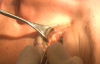
A desmarres retractor provides exposure and an infratarsal incision is created through conjunctiva and lower eyelid retractors. Ballotement of the globe assists in visualizing the fat pads and determining the proper incision location. Traction sutures placed in the proximal conjunctival edge aid in exposure. If exposure is inadequate, lateral canthotomy and inferior cantholysis may be required. Direct access is gained to the three lower eyelid fat pads without disruption of the orbital septum.
The orbital fat pads are debulked or mobilized as pedicles for repositioning to areas of concavity inferior to the orbital rim. Strict hemostasis is maintained with monopolar or bipolar cautery. The inferior oblique muscle is visualized and left undisturbed. Fat redraping can occur in the suborbicularis or subperiosteal plane after creating a pocket and releasing attachments. The fat pedicles are secured with percutaneous sutures or internal absorbable sutures. The suborbicularis oculi fat (SOOF) may be elevated and secured to the orbital rim periosteum with absorbable sutures via the transconjunctival incision. Similar to orbital fat repositioning, a SOOF lift aids in effacing the tear trough and infraorbital hollows.
The conjunctival incision may be approximated with absorbable sutures or may heal without direct closure.
Skin approach (infraciliary)
An incision is created 1-2 mm inferior to the eyelash line or within a preexisting infraciliary crease, extending to a lateral eyelid crease. A skin “pinch” may be used to determine the amount of redundancy by crushing the skin with a hemostat without causing traction on the eyelid margin. Alternatively, a skin flap may be created, extending as far as necessary to adequate mobilization without distortion of the shape of the eyelid aperture. A conservative amount of skin is removed to avoid anterior lamellar shortage. The patient is asked to gaze upward and open their mouth to assess the allowable amount of skin trim. The skin-muscle approach initiates a flap deep to the orbicularis and allows for superior advancement and trimming of skin and muscle individually or as a single unit. Access to the orbital fat pads and SOOF is possible from the infraciliary incision and is managed in the same way as the transconjunctival route. An infraciliary incison, either complete or lateral provides access to the orbicularis muscle and the orbitomalar ligament which can be elevated and suspended to the external lateral orbital rim periosteum in order to lift and support the eyelid. Lateral canthopexy can also be performed using the same incision and is often performed with infraciliary blepharoplasty to maintain or elevate the position of the lower eyelid.
The skin incision is closed with fine monofilament or absorbable catgut sutures.
Additional procedures
Significant lower lid laxity documented prior to blepharoplasty is managed with canthopexy or lateral canthoplasty.
Fat grafting techniques may be used to add volume to the infraorbital hollows and the lid-cheek junction. Alloplastic orbital rim and malar implants can also improve volume and projection deficiencies.
Laser skin ablative or non ablative resurfacing or chemical peels improve the lower eyelid skin quality and reduce rhytidosis and dyschromia in appropriate candidates.
Ligation, sclerotherapy, or laser treatment can reduce or eliminate the appearance of unwanted prominent lower eyelid veins.
Botulinum toxin injections minimize the dynamic creases that form in the periorbital region and lower lids.
Postoperative care
- Cold compresses are often recommended to reduce swelling in the first 48 hours, followed by warm compresses
- Bland ointment or ophthalmic antibiotic (or steroid/antibiotic combination) ointment or drops are applied for the first postoperative week
- Strenuous activity is avoided for a period of days or weeks after surgery
- A follow-up visit is planned within a week of surgery and non-absorbable sutures are removed 5 to 7 days after surgery to avoid suture tracks and excessive scarring
- Patients are instructed on concerning symptoms that would indicate bleeding or infection that would necessitate contact with their surgeon
Complications
- Retrobulbar hemorrhage is a rare but serious complication that should be emergently addressed
- Chemosis
- Pyogenic granuloma
- Undercorrection or overcorrection of steatoblepharon
- Lagophthalmos
- Inferior oblique muscle injury / diplopia
- Hypertrophic scar
- Suture cysts
- Lower eyelid retraction is a potential risk of lower blepharoplasty and may be more common when the septum is violated from the anterior approach as compared to the transconjunctival approach. Retraction is observed as a low-positioned eyelid that is tethered to the orbital rim due to scarring of the middle and/or posterior eyelid lamellae.
- Anterior lamellar shortage may also occur and is caused by overzealous skin removal, unfavorable contraction after surgery, or poor healing after eyelid skin resurfacing.
- Ectropion, independent of retraction or skin shortage can take place after lower blepharoplasty if a lax lower eyelid is left uncorrected or if postoperative tractional forces are unopposed in the setting of poor canthal support.
Additional Resources
References
- ↑ Baylis, H. I., Long, J. A., & Groth, M. J. (1989). Transconjunctival lower eyelid blepharoplasty: technique and complications. Ophthalmology, 96(7), 1027-1032.
- ↑ Wong, C. H., & Mendelson, B. (2017). Extended transconjunctival lower eyelid blepharoplasty with release of the tear trough ligament and fat redistribution. Plastic and reconstructive surgery, 140(2), 273-282.


