All content on Eyewiki is protected by copyright law and the Terms of Service. This content may not be reproduced, copied, or put into any artificial intelligence program, including large language and generative AI models, without permission from the Academy.
Disease entity
- ICD10- H21.509
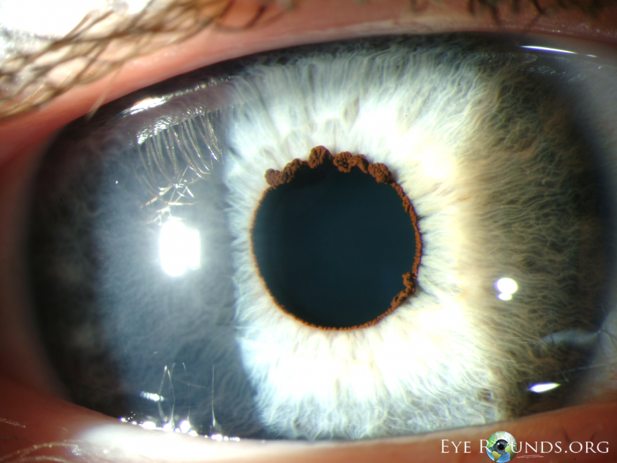
The word Flocculus is derived from the Latin floccus meaning tuft of wool. Flocculi are congenital, benign, cyst-like lesions present at the pupillary border of the iris. These tufts develop from the iris pigment epithelium and clinically present either as spherical or teardrop-shaped lesions.
Pathophysiology
Central iris pigmented epithelial cysts can undergo cycles of collapse and reformation, leading to the formation of wrinkled masses known as iris flocculi along the pupillary border. Despite their size, these lesions generally do not cause visual impairment. While iris flocculi are typically sporadic, they can also be familial and occasionally linked to aortic aneurysms and dissections.[1]
Etiopathogenesis
Iris flocculus is usually sporadic, however, familial variants have been described.
Iris flocculus has an association with specific genes like ACTA2 and MYH11. Mutations in the ACTA2 gene have an association with iris flocculi, familial TAAD (Thoracic aortic aneurysm and dissection)[2]. Iris sphincter muscles are made from smooth muscles. These muscles have a contraction-relaxation function assembly which is made up of actin and myosin filaments. Alpha actin 2 (ACTA-2 gene) regulates actin filaments function and MYH11 (Myosin heavy chain) controls myosin filaments.[3] These genes also control the smooth muscles of the aorta. Mutations in these 2 genes play a significant role in the development of aortic aneurysms (TAAD). ACTA-2 gene mutation accounts for 14% of familial TAAD[2] and it has also been associated with multisystemic smooth muscle dysfunction syndrome characterized by congenital mydriatic pupil with loss of accommodation and patent ductus arteriosus[4]. Disabella et al studied 100 TAAD patients and among them, only 6 patients had iris flocculi but they found that it's the most common extra aortic trait in TAAD.[5]
Role of ACTA2 gene
Mutation of ACTA2 gene causes impaired contraction of smooth muscle cell which can produce ophthalmic manifestation like iris flocculus and congenital mydriasis with impaired accommodation; and cardiac disease like nonsyndromic TAAD.[4]
Diagnosis
The patient is mostly asymptomatic and rarely has vision problems. On slit-lamp examination, these iris lesions present bilaterally like excrescent tufts overhanging on the pupillary zone. UBM and AS-OCT can be used to demonstrate finer details of these iris lesions.
Differential diagnosis
- Primary Cysts
- Iris pigment epithelium cysts
- Iris stromal cysts
- Secondary Cysts
- Epithelial downgrowth
- Post-surgical
- Post-traumatic
- Cysts secondary to intraocular tumors
- Iris melanoma
- Medulloepithelioma
- Iris pigment epithelium adenoma
- Medication-induced
- Miotics (phospholine iodide, pilocarpine)
- Prostaglandins including latanoprost
- Epithelial downgrowth
- Iris melanocytic tumours
- Iris freckle
- Iris nevus
- Iris melanocytoma
- Lisch nodule
- Iris and cilliary body melanoma
- Brushfield flecks
- Iris mammillations
- Parasite cyst
- Koeppe nodule
- Retinoblastoma
- Leiomyoma
- Xanthogranuloma
- Metastases from distant tumors
- Leukemic infiltrates
Clinical significance
Iris Flocculus is rare and not harmful but it can be a significant ocular marker for familial TAAD which is a life-threatening condition.
References
- ↑ Lewis RA, Merin LM. Iris flocculus and familial aortic dissection. Arch Ophthalmol 1995; 113(10):1330-1
- ↑ 2.0 2.1 Guo DC, Pannu H, Tran-Fadulu V, et al. Mutations in smooth muscle alpha-actin (ACTA2) lead to thoracic aortic aneurysms and dissections [published correction appears in Nat Genet. 2008 Feb;40(2):255]. Nat Genet. 2007;39(12):1488‐1493
- ↑ Risma TB, Alward WL. Successful long-term management of iris flocculi and miosis in a patient with a strong family history of thoracic aortic aneurysms and dissections associated with an MYH11 mutation. JAMA Ophthalmol. 2014;132(6):778‐780
- ↑ 4.0 4.1 Milewics DM, Ostergaard JR, Ala-Kokko LM, et al. De novo ACTA2 mutation causes a novel syndrome of multisystemic smooth muscle dysfunction. Am J Med Genet. 2010;152A:2437–43
- ↑ Disabella E, Grasso M, Gambarin FI. Risk of dissection in thoracic aneurysms associated with mutations of smooth muscle alpha-actin 2 (ACTA2). Heart. 2011;97(4):321‐326.


