File:Vitreoretinal interface bonds.jpg
Original file (1,211 × 314 pixels, file size: 190 KB, MIME type: image/jpeg)
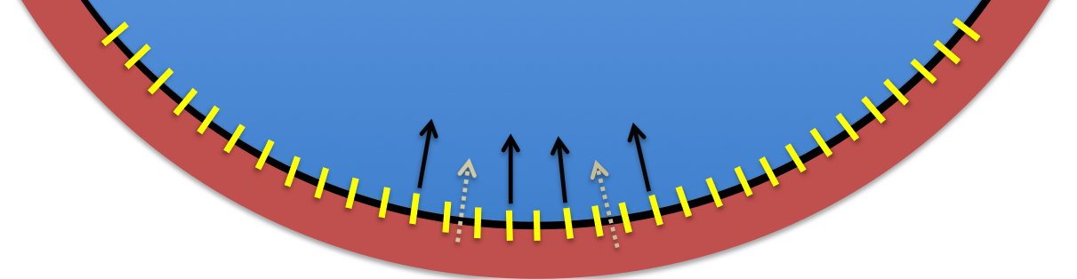
Pictorial representation of vitreoretinal interface and tractional forces active in vitreomacular traction: Solid black arrows represent posterior to anterior forces generated by the vitreous due to liquefaction and condensation.
Yellow lines represent vitreoretinal interface bonds constituted by laminin, fibronectin, and collagen types VI, VII, XVIII. These also represent the sites of action of ocriplasmin used for pharmacologic vitreolysis in vitreomacular traction syndrome.
Broken white arrows represent tractional forces transmitted to the retina via the above vitreoretinal bonds/attachments. These forces can eventually produce vitreomacular traction syndrome.
File history
Click on a date/time to view the file as it appeared at that time.
| Date/Time | Thumbnail | Dimensions | User | Comment | |
|---|---|---|---|---|---|
| current | 13:45, December 1, 2013 | 1,211 × 314 (190 KB) | Sanket.U.Shah (talk | contribs) | Pictorial representation of vitreoretinal interface and tractional forces active in vitreomacular traction: Solid black arrows represent posterior to anterior forces generated by the vitreous due to liquefaction and condensation. Yellow lines represent v |
You cannot overwrite this file.
File usage
The following page uses this file: