File:Serpiginous chorioretinopathy.png
Serpiginous_chorioretinopathy.png (589 × 527 pixels, file size: 209 KB, MIME type: image/png)
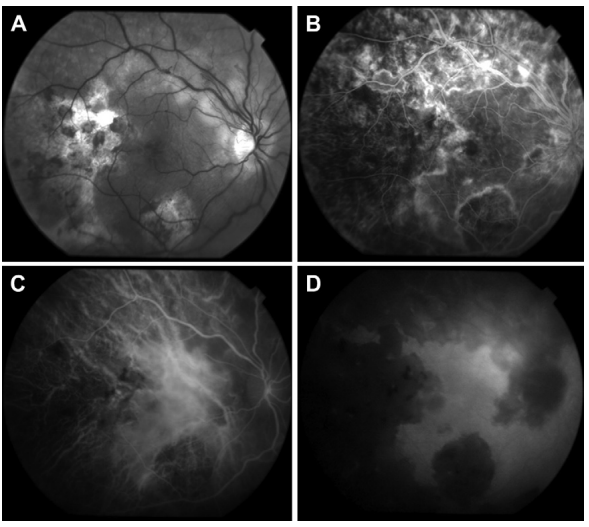
Serpiginous Choroiditis: 34-year old Afghani woman with history of bilateral choroiditis. A: Red free fundus photography of the right eye reveals geographic areas of choroidal atrophy, pigment clumping, and cystoid macular edema. B: In FA, border staining delineates atrophic areas with some blockage from pigment proliferation. Early accumulation of dye in cystoid spaces is evident. C, D: Mid-phase and late phase indocyanine green angiography reveals choriocapillaris loss in areas of healed lesions with preservation of large choroidal vessels. Choroidal circulation in non-involved areas is normal. Nazari Khanamiri H, Rao NA. Serpiginous choroiditis and infectious multifocal serpiginoid choroiditis. Surv Ophthalmol. 2013;58(3):203-232. doi:10.1016/j.survophthal.2012.08.008; Permission granted by Hossein Nazari MD PhD
File history
Click on a date/time to view the file as it appeared at that time.
| Date/Time | Thumbnail | Dimensions | User | Comment | |
|---|---|---|---|---|---|
| current | 18:04, October 22, 2023 | | 589 × 527 (209 KB) | Evan.Wotipka (talk | contribs) |
You cannot overwrite this file.
File usage
There are no pages that use this file.