File:Progression of Dry AMD Over 76 Months.jpg
From EyeWiki
Size of this preview: 800 × 284 pixels. Other resolution: 927 × 329 pixels.
Original file (927 × 329 pixels, file size: 70 KB, MIME type: image/jpeg)
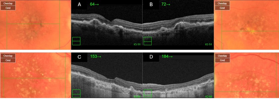
Baseline OCT images of the right eye (A) and left eye (B) show the initial state of drusenoid PEDs and pigmentary changes in dry AMD. After 76 months (C and D), the images reveal the development of geographic atrophy, the disappearance of drusenoid PEDs, and notable pigmentary alterations. (Courtesy of J. Khadamy)
File history
Click on a date/time to view the file as it appeared at that time.
| Date/Time | Thumbnail | Dimensions | User | Comment | |
|---|---|---|---|---|---|
| current | 06:00, July 26, 2024 | 927 × 329 (70 KB) | Joobin.Khadamy (talk | contribs) |
You cannot overwrite this file.
File usage
The following page uses this file: