File:Picture 3 OCTA.png
From EyeWiki

Size of this preview: 800 × 370 pixels. Other resolution: 1,214 × 561 pixels.
Original file (1,214 × 561 pixels, file size: 899 KB, MIME type: image/png)
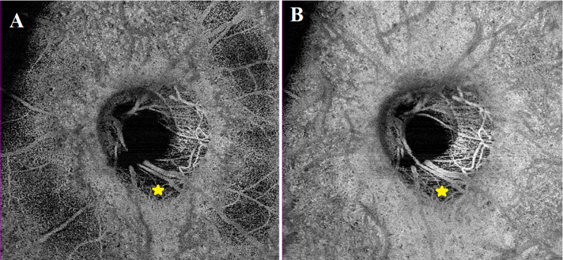
Picture 3: Same patient as in pictures 1 and 2. OCTA shots at the level of the choriocapillaris (A) and the choroid (B); 6x6 mm field of view centered on the papilla. The PICC appears as a hyporeflective area (yellow star), devoid of vascularization. The device used is the PLEX Elite® 9000 SS OCTA (Carl Zeiss Meditec AG, Jena, Germany).
File history
Click on a date/time to view the file as it appeared at that time.
| Date/Time | Thumbnail | Dimensions | User | Comment | |
|---|---|---|---|---|---|
| current | 11:53, April 28, 2022 | | 1,214 × 561 (899 KB) | Artemise.Dugauquier (talk | contribs) |
You cannot overwrite this file.
File usage
The following page uses this file: