File:Picture 2 OCT.png
From EyeWiki
Size of this preview: 800 × 596 pixels. Other resolution: 947 × 705 pixels.
Original file (947 × 705 pixels, file size: 632 KB, MIME type: image/png)
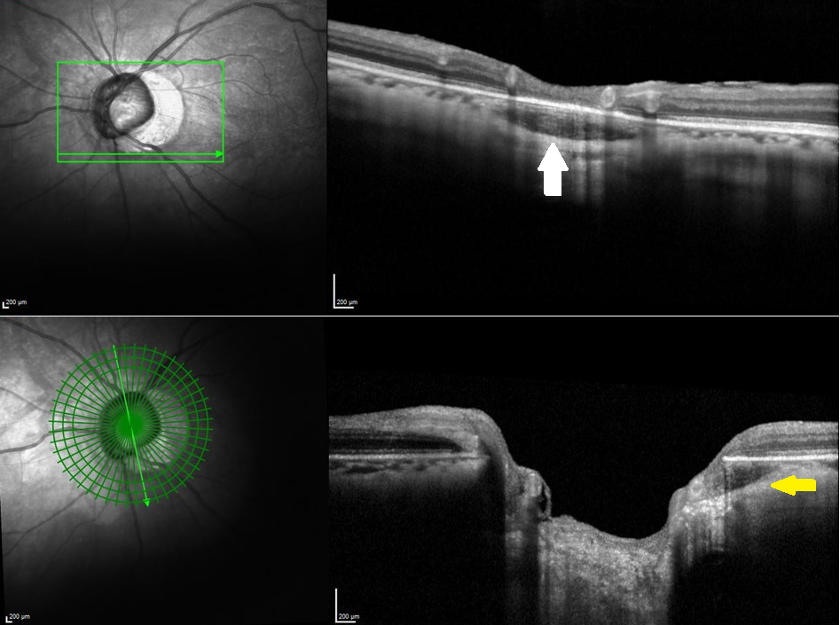
Same patient as in picture 1. Up. A horizontal linear OCT scan across the peripapillary region demonstrates a hyporeflective space (white arrow) under the intact retinal pigmentary epithelium. Down. On a radial scan, the PICC is seen as a choroidal hyporeflective space as well (yellow arrow); border tissue is absent. OCTs were made using the Spectralis® model S3300 (Heidelberg Engineering GmbH, Heidelberg, Germany).
File history
Click on a date/time to view the file as it appeared at that time.
| Date/Time | Thumbnail | Dimensions | User | Comment | |
|---|---|---|---|---|---|
| current | 11:49, April 28, 2022 | | 947 × 705 (632 KB) | Artemise.Dugauquier (talk | contribs) |
You cannot overwrite this file.
File usage
The following page uses this file: