File:Ocular Rosacea.png

Original file (3,000 × 4,339 pixels, file size: 4.78 MB, MIME type: image/png)
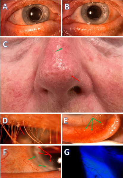
Cutaneous and Ocular Manifestations in a Rosacea Patient: A and B: The eyelid margins are rounded, notched, thickened, and exhibit telangiectasia. C: Phymatous rosacea presents on the nose (rhinophyma) and cheeks, marked by skin thickening, irregular nodular surface, telangiectasia (green arrow), fibrosis, and increased sebaceous gland volume (red arrow). D: Anterior blepharitis with waxy collarettes (red arrow) at the eyelash base. E: Conjunctival concretions (green arrows) on the palpebral conjunctiva indicate chronic inflammation. F: Abnormal blood vessel ingrowth (green arrows) and peripheral corneal scarring (red arrows), suggesting previous marginal keratitis episodes. G: Bulbar conjunctival staining in the lower areas adjacent to the eyelid margins. (Courtesy of J Khadamy)
File history
Click on a date/time to view the file as it appeared at that time.
| Date/Time | Thumbnail | Dimensions | User | Comment | |
|---|---|---|---|---|---|
| current | 02:30, August 3, 2024 | | 3,000 × 4,339 (4.78 MB) | Joobin.Khadamy (talk | contribs) |
You cannot overwrite this file.
File usage
The following page uses this file: