File:OCT scan and retinal layers.png
From EyeWiki
Size of this preview: 800 × 423 pixels. Other resolution: 1,596 × 843 pixels.
Original file (1,596 × 843 pixels, file size: 1.13 MB, MIME type: image/png)
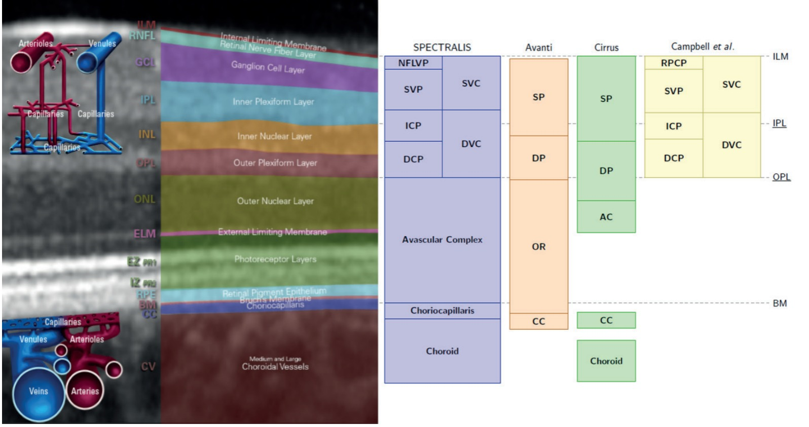
Left- B-scan OCT image with schematic representation of retinal and choroidal vascularization and identification of the respective layers. Right- Correspondence of segmentation schemes of different OCT-A devices.
Figure originally published in: SPECTRALIS Optical Coherence Tomography Angiography: Principles and Clinical Applications | Heidelberg Engineering GmbH, 2020 (5).
File history
Click on a date/time to view the file as it appeared at that time.
| Date/Time | Thumbnail | Dimensions | User | Comment | |
|---|---|---|---|---|---|
| current | 19:08, November 29, 2022 | | 1,596 × 843 (1.13 MB) | Koushik.Tripathy (talk | contribs) | Higher resolution |
| 02:44, April 20, 2022 | 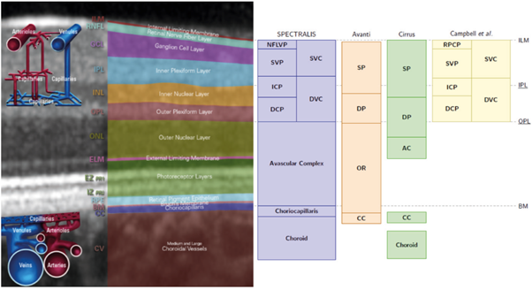 | 602 × 328 (228 KB) | Rita.Basto (talk | contribs) |
You cannot overwrite this file.
File usage
The following page uses this file: