File:Meesman-3.jpg
From EyeWiki

No higher resolution available.
Meesman-3.jpg (600 × 478 pixels, file size: 21 KB, MIME type: image/jpeg)
Meesmann Corneal Dystrophy. Slit-lamp photography demonstrates diffuse minute bubble-like intraepithelial microcysts.
Venckus T. Meesmann Corneal Dystrophy. Meesmann Epithelial Corneal Dystrophy. https://webeye.ophth.uiowa.edu/eyeforum/atlas/pages/Meesmann-epithelial-corneal-dystrophy/index.htm. Published January 2015. Accessed June 1, 2020.
File history
Click on a date/time to view the file as it appeared at that time.
| Date/Time | Thumbnail | Dimensions | User | Comment | |
|---|---|---|---|---|---|
| current | 16:22, June 1, 2020 | | 600 × 478 (21 KB) | Marianna.Kavalaraki (talk | contribs) | Meesmann Corneal Dystrophy. Slit-lamp photography demonstrates diffuse minute bubble-like intraepithelial microcysts. Contributor : Christopher Kirkpatrick. Eye Rounds Org. University of Iowa |
| 15:57, June 1, 2020 | 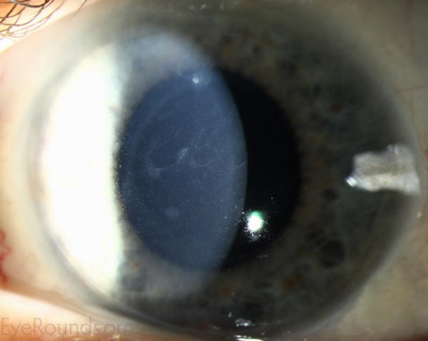 | 600 × 478 (21 KB) | Marianna.Kavalaraki (talk | contribs) | Meesmann Corneal Dystrophy. Slit-lamp photography demonstrates diffuse minute bubble-like intraepithelial microcysts. Venckus T. Meesmann Corneal Dystrophy. Meesmann Epithelial Corneal Dystrophy. https://webeye.ophth.uiowa.edu/eyeforum/atlas/pages/M... |
You cannot overwrite this file.
File usage
The following page uses this file: