File:Figure 1C.jpg
From EyeWiki

Size of this preview: 800 × 503 pixels. Other resolution: 3,007 × 1,892 pixels.
Original file (3,007 × 1,892 pixels, file size: 488 KB, MIME type: image/jpeg)
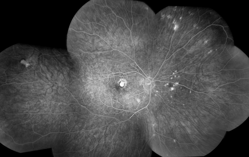
Late phase of the angiogram shows leakage from the CNV along with multiple punctate hyperfluorescent lesions in the peripapillary area and the periphery.
Courtesy of Dr Narsing Rao and taken from the following publication: Pachydaki SI, Jakobiec FA, Bhat P, Sobrin L, Michaud NA, Seshan SV, D'Amico DJ. Surgical management and ultrastructural study of choroidal neovascularization in punctate inner choroidopathy after bevacizumab. J Ophthalmic Inflamm Infect. 2011 Nov 27.
File history
Click on a date/time to view the file as it appeared at that time.
| Date/Time | Thumbnail | Dimensions | User | Comment | |
|---|---|---|---|---|---|
| current | 21:32, November 30, 2011 | | 3,007 × 1,892 (488 KB) | Katrina.A.Mears (talk | contribs) | Late phase of the angiogram shows leakage from the CNV along with multiple punctate hyperfluorescent lesions in the peripapillary area and the periphery. Courtesy of Dr Narsing Rao and taken from the following publication: Pachydaki SI, Jakobiec FA, Bh |
You cannot overwrite this file.
File usage
There are no pages that use this file.