File:Figure 1.jpg
From EyeWiki
Size of this preview: 586 × 600 pixels. Other resolution: 1,889 × 1,934 pixels.
Original file (1,889 × 1,934 pixels, file size: 391 KB, MIME type: image/jpeg)
Figure 1. Lifting of LASIK flap.
File history
Click on a date/time to view the file as it appeared at that time.
| Date/Time | Thumbnail | Dimensions | User | Comment | |
|---|---|---|---|---|---|
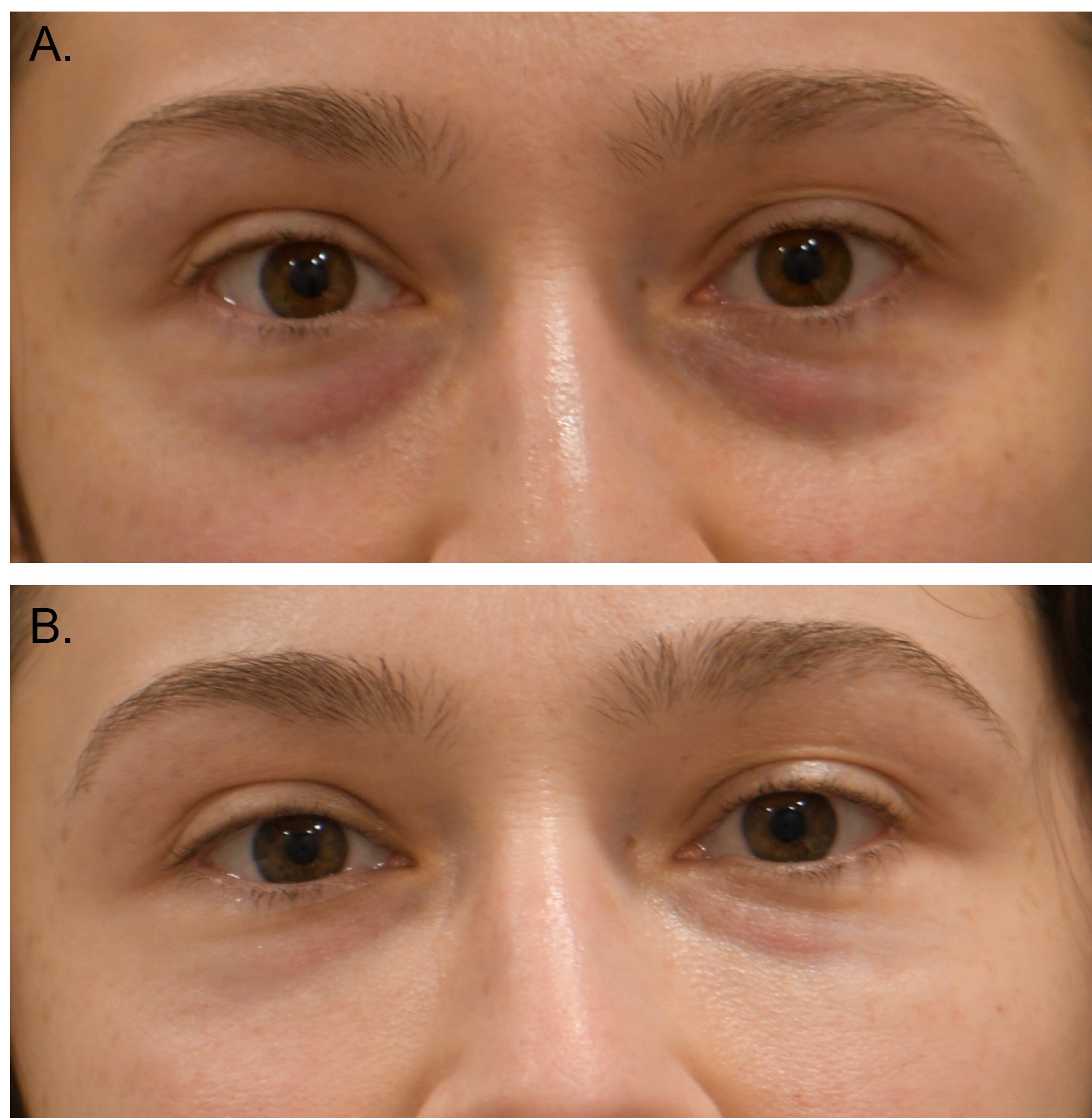
| current | 16:13, June 20, 2023 | | 1,889 × 1,934 (391 KB) | Jan.Ulloa (talk | contribs) | Figure 1: Tear through deformity treated with filler. A, Pre-filler injection. B, Post-filler injection. |
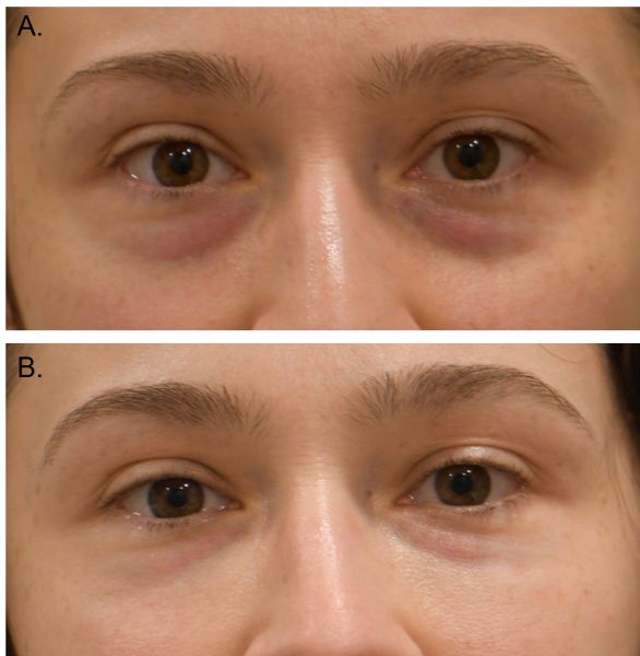
| 19:35, June 6, 2019 | 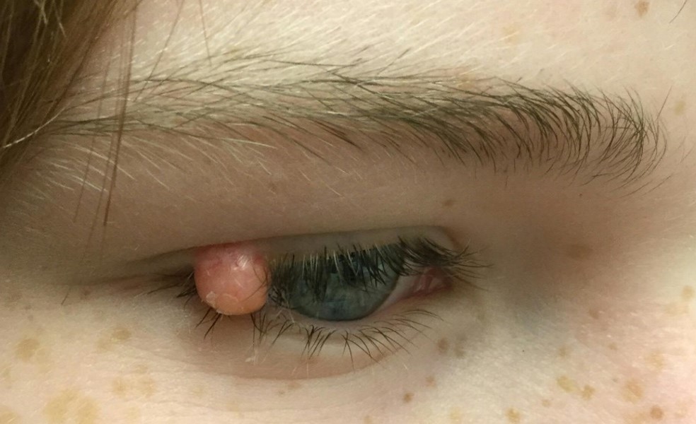 | 984 × 600 (102 KB) | Sarah.Xu (talk | contribs) | JXG figure 1 | |
| 09:12, December 1, 2016 | 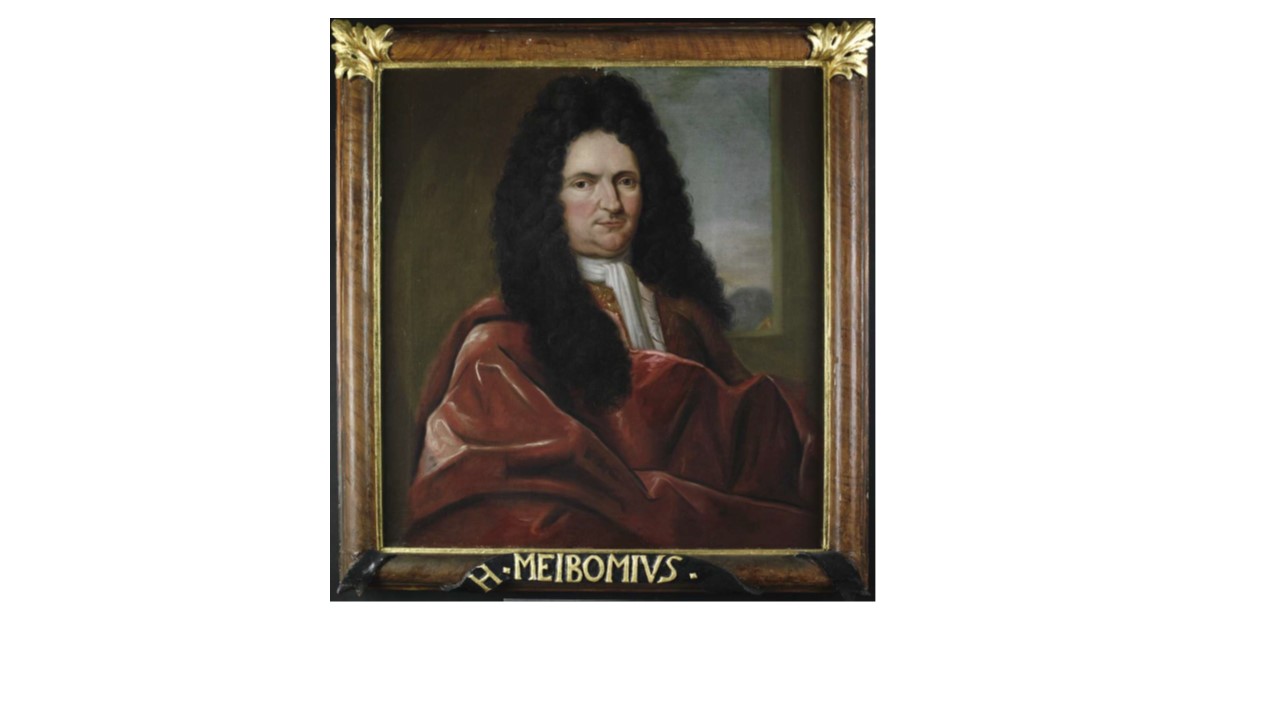 | 1,280 × 720 (73 KB) | Deepak.Sambhara (talk | contribs) | Figure 1. Dr. Heinrich Meibom. Reprinted with permission of the Herzog August Bibliothek, Wolfenbu¨ttel, Germany, Signatur B 100. | |
| 06:49, April 1, 2013 | 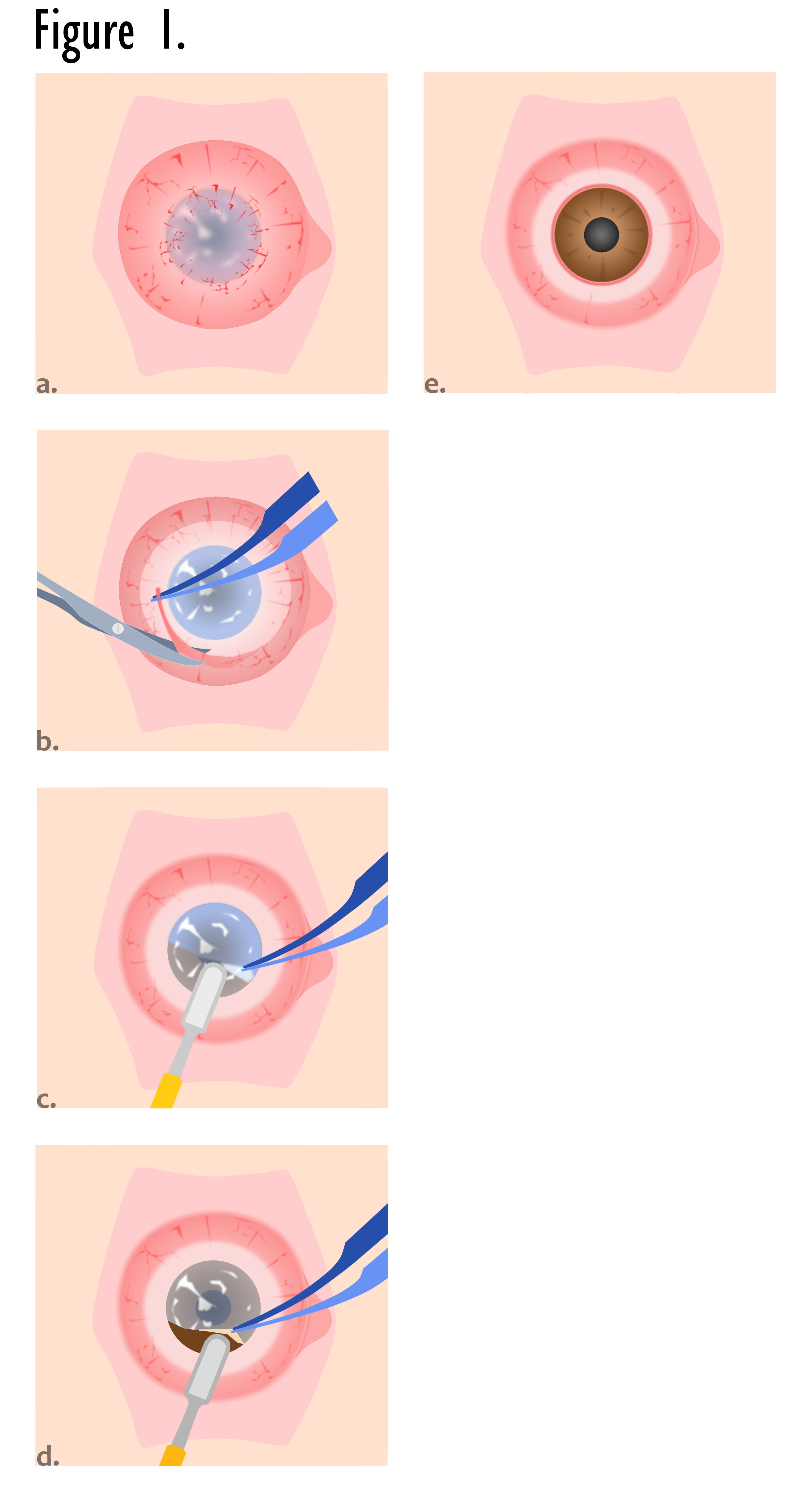 | 2,721 × 5,020 (595 KB) | Seyed-Farzad (talk | contribs) | Bed preparation; a. vascularized stem cell deficient external eye surface and corneal opacity (conjunctival shortage and scarring is not shown); b. dissection and removal of fibrovascular scar; c. & d. lamellar keratectomy; e. clear deep cornea of the rec | |
| 23:04, November 25, 2011 | 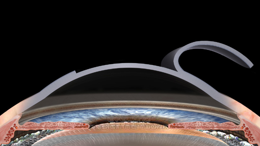 | 840 × 472 (79 KB) | Lespandar (talk | contribs) | Reverted to version as of 19:53, 22 June 2010 | |
| 23:03, November 25, 2011 |  | 200 × 198 (10 KB) | Lespandar (talk | contribs) | ||
| 12:53, June 22, 2010 |  | 840 × 472 (79 KB) | Davidhuang (talk | contribs) | Figure 1. Lifting of LASIK flap. |
You cannot overwrite this file.
File usage
The following file is a duplicate of this file (more details):
There are no pages that use this file.