File:Fig2.png
From EyeWiki
Size of this preview: 800 × 312 pixels. Other resolution: 911 × 355 pixels.
Original file (911 × 355 pixels, file size: 418 KB, MIME type: image/png)
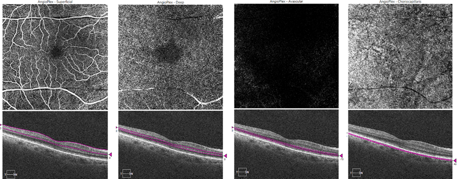
En-face angiograms (and representation of the respective plane in B-scans) at the level of the superficial plexus (a), deep plexus (b), avascular retina (c) and choriocapillary layer (d).
Images acquired with the OCT-CIRRUS AngioPlex™ system (Carl Zeiss Meditec Inc., Dublin, CA, USA).
File history
Click on a date/time to view the file as it appeared at that time.
| Date/Time | Thumbnail | Dimensions | User | Comment | |
|---|---|---|---|---|---|
| current | 02:41, April 20, 2022 | 911 × 355 (418 KB) | Rita.Basto (talk | contribs) |
You cannot overwrite this file.
File usage
The following page uses this file: