File:Fig.14 avoid suprachor.png
From EyeWiki
Size of this preview: 800 × 347 pixels. Other resolution: 892 × 387 pixels.
Original file (892 × 387 pixels, file size: 598 KB, MIME type: image/png)
Summary
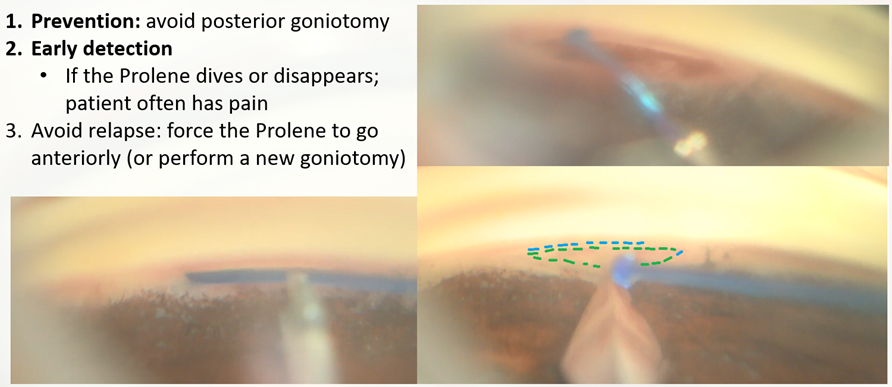
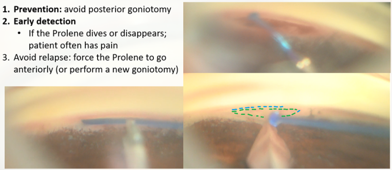
Figure 14. Avoiding the suprachoroidal path. On the left side of the image, a supra-choroidal path is identified; this was facilitated by a posterior goniotomy (on the image bottom right, the green strokes show the goniotomy performed, while the blue strokes show the ideal goniotomy position). We withdrew the suture, injected abundant OVD (there was significant bleeding), conducted a more anterior goniotomy, and then inserted the suture with a more anterior bias. The suture went successfully into the Schlemm’s canal.
File history
Click on a date/time to view the file as it appeared at that time.
| Date/Time | Thumbnail | Dimensions | User | Comment | |
|---|---|---|---|---|---|
| current | 09:51, May 29, 2023 | | 892 × 387 (598 KB) | Ana.Miguel (talk | contribs) | Figure 14. Avoiding the suprachoroidal path. On the left side of the image, a supra-choroidal path is identified; this was facilitated by a posterior goniotomy (on the image bottom right, the green strokes show the goniotomy performed, while the blue strokes show the ideal goniotomy position). We withdrew the suture, injected abundant OVD (there was significant bleeding), conducted a more anterior goniotomy, and then inserted the suture with a more anterior bias. The suture went successfully... |
You cannot overwrite this file.
File usage
The following page uses this file: