File:Fig.11 eatt.png
From EyeWiki

Size of this preview: 588 × 599 pixels. Other resolution: 977 × 996 pixels.
Original file (977 × 996 pixels, file size: 1.17 MB, MIME type: image/png)
Summary
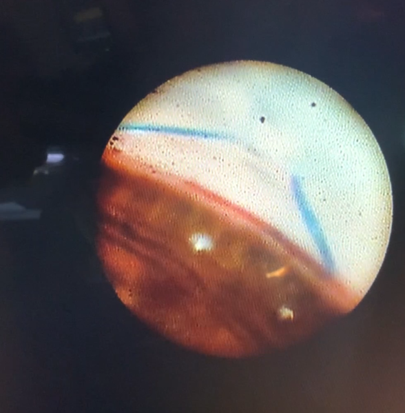
Figure 11. EATT procedure. This endoscopic camera allows a visualization from the inside out; in the figure, there is a depiction of the final stage of the GATT: a trabeculotomy, with the suture being removed from the eye by the main incision (center of the image) and causing a trabeculotomy (right of the image).
File history
Click on a date/time to view the file as it appeared at that time.
| Date/Time | Thumbnail | Dimensions | User | Comment | |
|---|---|---|---|---|---|
| current | 09:50, May 29, 2023 | | 977 × 996 (1.17 MB) | Ana.Miguel (talk | contribs) | Figure 11. EATT procedure. This endoscopic camera allows a visualization from the inside out; in the figure, there is a depiction of the final stage of the GATT: a trabeculotomy, with the suture being removed from the eye by the main incision (center of the image) and causing a trabeculotomy (right of the image). |
You cannot overwrite this file.
File usage
The following page uses this file: