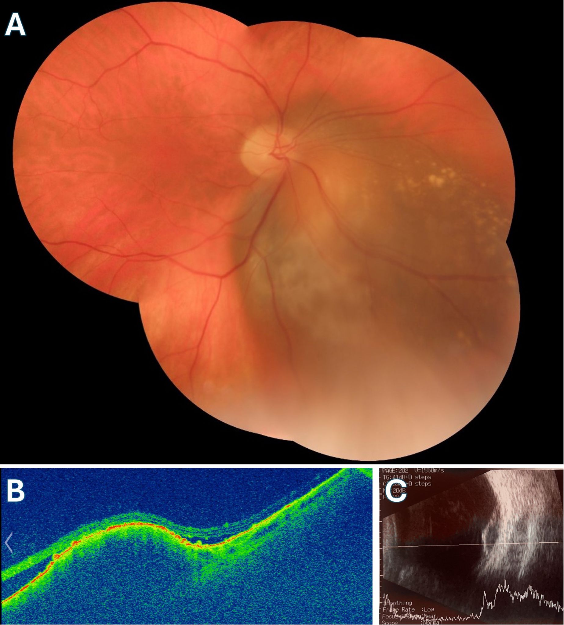
File:Choroidal Melanoma.jpg

Original file (4,092 × 4,537 pixels, file size: 1.79 MB, MIME type: image/jpeg)
A: Mosaic fundus image displaying a peripapillary choroidal melanoma measuring 6x5 disc diameters with a thickness of 2 mm. The image reveals suspected orange pigment, drusen, and the presence of subretinal fluid. B: Widefield OCT B-scan showing a choroidal elevation with notable choroidal shadowing. Subretinal fluid (SRF) is visible on the sides of the elevation, along with subretinal hyperreflective material (SRHM) and drusen. C: Ultrasonography image depicting the choroidal elevation with low internal echogenicity, characteristic of choroidal melanoma. (Courtesy of J. Khadamy)
File history
Click on a date/time to view the file as it appeared at that time.
| Date/Time | Thumbnail | Dimensions | User | Comment | |
|---|---|---|---|---|---|
| current | 08:15, August 31, 2024 | | 4,092 × 4,537 (1.79 MB) | Joobin.Khadamy (talk | contribs) |
You cannot overwrite this file.
File usage
The following page uses this file: