File:CNV Type 1.jpg
From EyeWiki
No higher resolution available.
CNV_Type_1.jpg (720 × 392 pixels, file size: 90 KB, MIME type: image/jpeg)
Summary
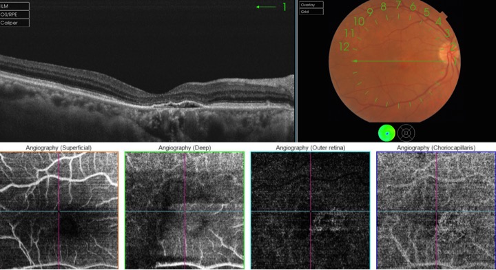
Type 1 CNV. Type 1 neovascular lesion is located below the RPE as seen in the B-scan frame. In the OCT-A frame, a neovascular coralliform network is observed which emanates from the choroidal vasculature and extends to the sub-RPE space. (Courtesy of Andrea Tamine Hoyos Dumar, MD)
File history
Click on a date/time to view the file as it appeared at that time.
| Date/Time | Thumbnail | Dimensions | User | Comment | |
|---|---|---|---|---|---|
| current | 00:47, June 3, 2017 | | 720 × 392 (90 KB) | Dan.Mummert.AAO (talk | contribs) |
You cannot overwrite this file.
File usage
The following page uses this file: