File:BRVO.PNG
From EyeWiki

Size of this preview: 696 × 599 pixels. Other resolution: 733 × 631 pixels.
Original file (733 × 631 pixels, file size: 852 KB, MIME type: image/png)
Creative common license: http://creativecommons.org/licenses/by-nc/2.0/ ; http://creativecommons.org/licenses/by-nc/2.0/legalcode
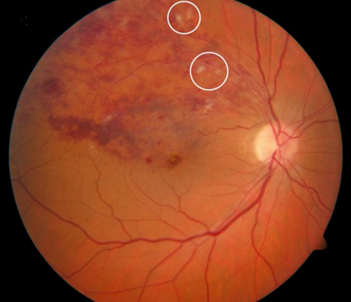
Branch retinal vein occlusion of superotemporal branch vein in the right eye. Fundus photograph showing widespread haemorrhages and axonal congestion (cotton wool spots; white circles) upstream of the venous occlusion.
File history
Click on a date/time to view the file as it appeared at that time.
| Date/Time | Thumbnail | Dimensions | User | Comment | |
|---|---|---|---|---|---|
| current | 11:22, November 30, 2015 | | 733 × 631 (852 KB) | Musa.Abdelaziz (talk | contribs) | Creative common license: http://creativecommons.org/licenses/by-nc/2.0/ ; http://creativecommons.org/licenses/by-nc/2.0/legalcode Branch retinal vein occlusion of superotemporal branch vein in the right eye. Fundus photograph showing widespread haemo... |
You cannot overwrite this file.
File usage
The following page uses this file: