File:Advanced AMD progression-.png
Advanced_AMD_progression-.png (742 × 215 pixels, file size: 239 KB, MIME type: image/png)
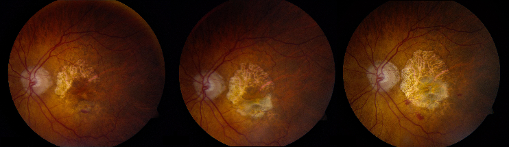
Time-lapse fundus photography over the course of 29 months showing progression of geographic atrophy in a patient with advanced ARMD. The photograph demonstrates a central residual island of retinal pigment epithelium in the first frame (visual acuity 20/40) that undergoes atrophy by the second frame (8 months later, visual acuity 20/150), and progressively more atrophy 21 months later in the third frame(visual acuity 20/400 eccentric). The inferior macula demonstrates some exudative changes (subretinal fibrosis and hemorrhage).
File history
Click on a date/time to view the file as it appeared at that time.
| Date/Time | Thumbnail | Dimensions | User | Comment | |
|---|---|---|---|---|---|
| current | 17:00, May 24, 2012 | 742 × 215 (239 KB) | Yasser.M.Elshatory (talk | contribs) | Time-lapse fundus photography over the course of 29 months showing progression of geographic atrophy in a patient with advanced ARMD. The photograph demonstrates a central residual island of retinal pigment epithelium in the first frame (visual acuity 20 |
You cannot overwrite this file.
File usage
The following page uses this file: