File:APMPPE OCTA.jpg
APMPPE_OCTA.jpg (716 × 274 pixels, file size: 119 KB, MIME type: image/jpeg)
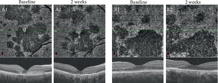
OCT-A: flow detected at the level of the choriocapillaris is disrupted in active lesions (baseline) and can improve with time or treatment in patients with APMPPE (left panel: OD; right panel: OS). (Copyright © 2020 Mariana A. Oliveira et al. Reproduced without modifications from an open access article, Management of Acute Posterior Multifocal Placoid Pigment Epitheliopathy (APMPPE): Insights from Multimodal Imaging with OCTA, distributed under the Creative Commons Attribution License; https://www.ncbi.nlm.nih.gov/pmc/articles/PMC7094199/)
File history
Click on a date/time to view the file as it appeared at that time.
| Date/Time | Thumbnail | Dimensions | User | Comment | |
|---|---|---|---|---|---|
| current | 15:03, August 8, 2020 | 716 × 274 (119 KB) | Michael.Massengill (talk | contribs) | OCT-A: flow detected at the level of the choriocapillaris is disrupted in active lesions (baseline) and can improve with time or treatment in patients with APMPPE (left panel: OD; right panel: OS). (Copyright © 2020 Mariana A. Oliveira et al. Reproduc... |
You cannot overwrite this file.
File usage
The following page uses this file: