File:AA0 OCTA.jpg
From EyeWiki
Size of this preview: 292 × 598 pixels. Other resolution: 1,125 × 2,305 pixels.
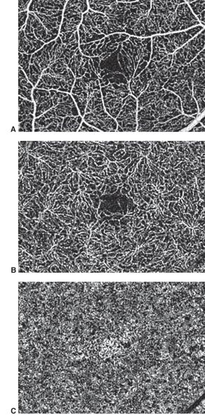
Original file (1,125 × 2,305 pixels, file size: 468 KB, MIME type: image/jpeg)
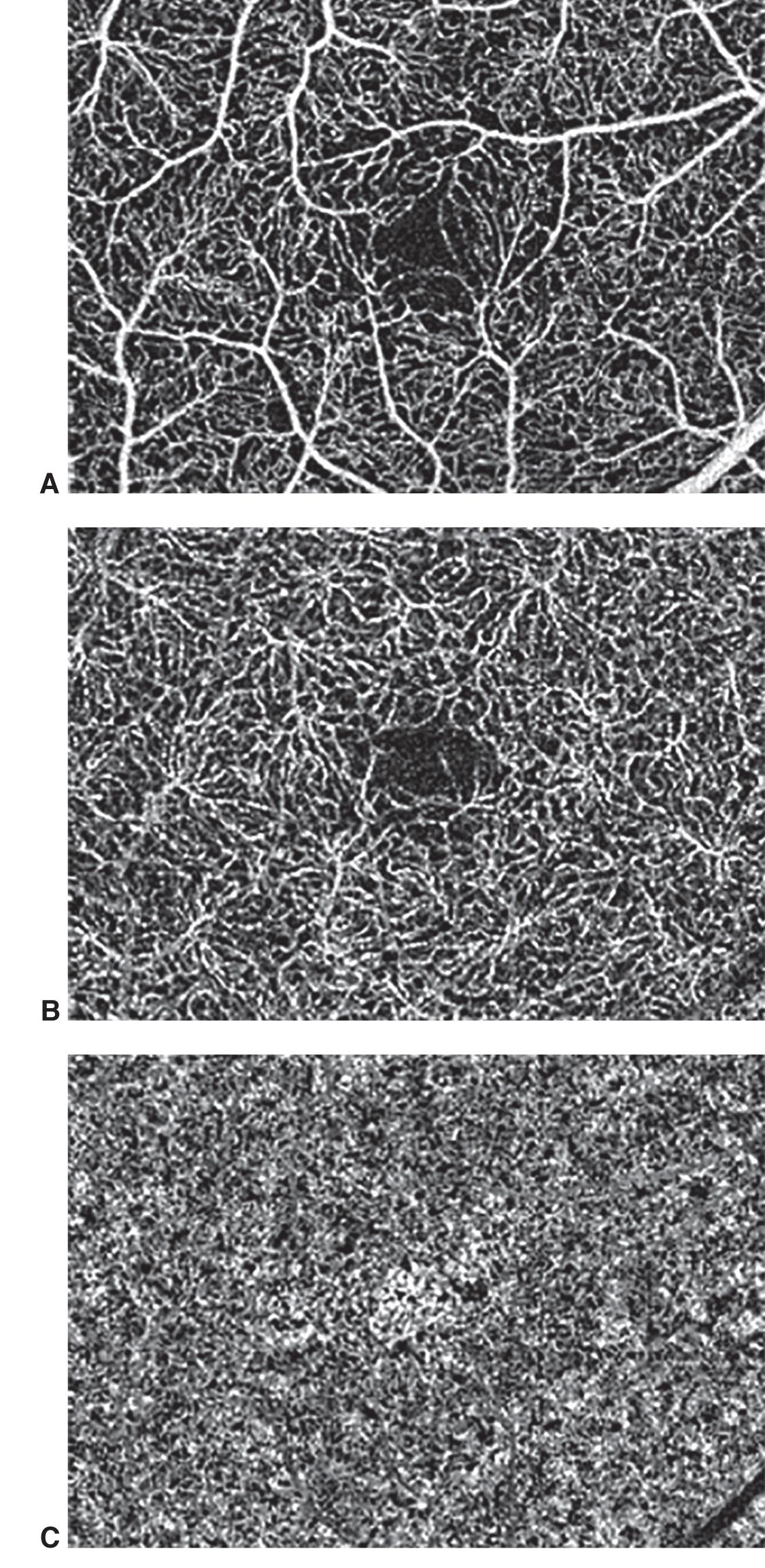
Courtesy of Richard F. Spaide, MD. OCT angiography with projection artifact removal. A, Image showing a superficial vascular plexus with fractal branching. B, Image showing a deep vascular plexus. Its vessels are small and do not show the same branching characteristic as the superficial vascular plexus. C, Image of the choriocapillaris with dark areas; these low-signal areas are called signal voids.
File history
Click on a date/time to view the file as it appeared at that time.
| Date/Time | Thumbnail | Dimensions | User | Comment | |
|---|---|---|---|---|---|
| current | 20:52, October 2, 2023 | | 1,125 × 2,305 (468 KB) | Claudia.Prospero.Ponce (talk | contribs) |
You cannot overwrite this file.
File usage
The following page uses this file: