File:AA0 53575.jpg
From EyeWiki
Size of this preview: 373 × 598 pixels. Other resolution: 1,125 × 1,805 pixels.
Original file (1,125 × 1,805 pixels, file size: 201 KB, MIME type: image/jpeg)
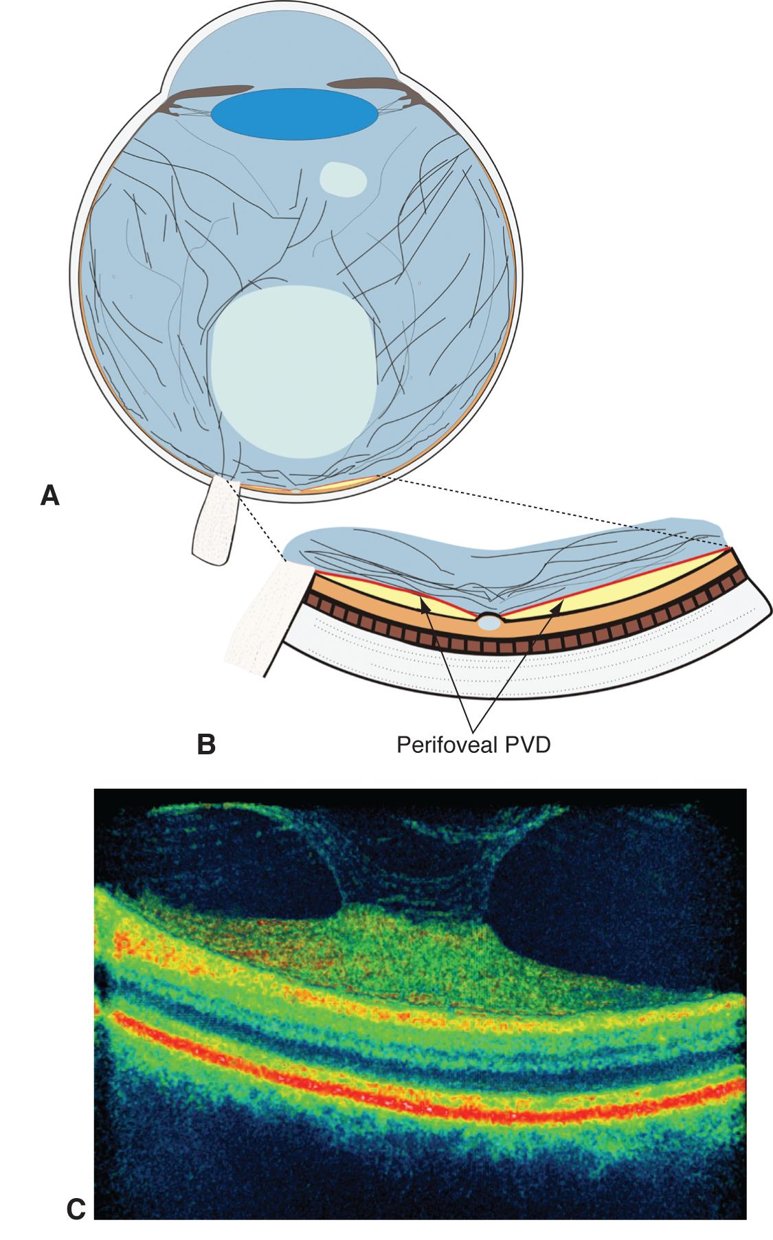
Pictorial representation of the eye shows two pockets of vitreous gel liquefaction (A) and a perifoveal posterior vitreous detachment associated with persistent attachment of the vitreous to the foveal region (B). Optical coherence tomography of the vitreomacular interface shows vitreomacular traction (C) resulting from the incomplete posterior vitreous detachment.
(c) 2013 American Academy of Ophthalmology
File history
Click on a date/time to view the file as it appeared at that time.
| Date/Time | Thumbnail | Dimensions | User | Comment | |
|---|---|---|---|---|---|
| current | 21:24, November 15, 2013 | | 1,125 × 1,805 (201 KB) | Sanket.U.Shah (talk | contribs) | Pictorial representation of the eye shows two pockets of vitreous gel liquefaction (A) and a perifoveal posterior vitreous detachment associated with persistent attachment of the vitreous to the foveal region (B). Optical coherence tomography of the vitre |
You cannot overwrite this file.
File usage
The following page uses this file: