File:3.jpg
From EyeWiki

Size of this preview: 612 × 600 pixels. Other resolution: 888 × 870 pixels.
Original file (888 × 870 pixels, file size: 155 KB, MIME type: image/jpeg)
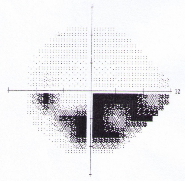
This is standard automated perimetry of the left eye of a patient with POAG. Note the dense inferior arcuate scotoma which respects the horizontal meridian, and is denser nasally.
File history
Click on a date/time to view the file as it appeared at that time.
| Date/Time | Thumbnail | Dimensions | User | Comment | |
|---|---|---|---|---|---|
| current | 19:05, June 14, 2010 | | 888 × 870 (155 KB) | Anthony.P.Khawaja.CMT (talk | contribs) | This is standard automated perimetry of the left eye of a patient with POAG. Note the dense inferior arcuate scotoma which respects the horizontal meridian, and is denser nasally. |
You cannot overwrite this file.
File usage
The following page uses this file: