All content on Eyewiki is protected by copyright law and the Terms of Service. This content may not be reproduced, copied, or put into any artificial intelligence program, including large language and generative AI models, without permission from the Academy.
Disease Entity
Strabismus/ocular misalignment
Disease
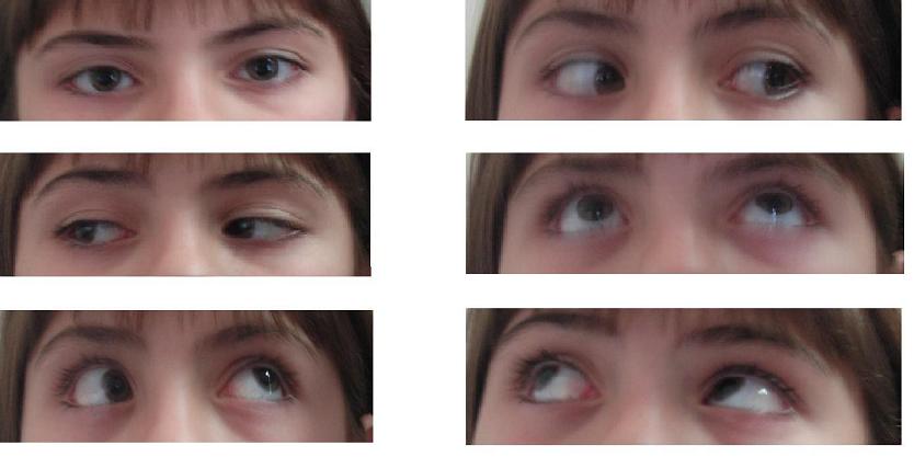
Brown syndrome is a form of vertical strabismus characterized by limited elevation of the eye in an adducted position, most often secondary to mechanical restriction of the superior oblique tendon-trochlea complex.
Brown syndrome can be congenital or acquired, is bilateral in approximately 10% of cases, and has a slight predilection for females. There is thought to be a genetic predisposition to congenital Brown syndrome, however, most cases are sporadic in nature.
Based on the 9-gaze pattern, it can be confused for an inferior oblique palsy. Differentiation between IO palsy and SO restriction of Brown’s can be done using Forced Duction Test. As it is a painful test, it is difficult to perform in children without general anesthesia.
Pathophysiology and Etiology
There is ongoing controversy regarding the possible underlying reasons for Brown Syndrome.
Previously referred to as "superior oblique tendon-sheath syndrome," Brown Syndrome was first described by Dr. Harold Whaley Brown in 1950.[1] It was initially considered a dysgenesis of the superior oblique muscle's tendon sheath.[2] More recent evidence suggests the characteristic restriction of elevation in adduction may be due to abnormalities of the tendon-trochlea complex.[3] Other studies propose anomalies in the development of the extraocular muscles nervous system, such as congenital cranial dysinnervation disorders (CCDDs), as a possible reason in some cases.[4] A newer explanation refers to the presence of a fibrotic strand found in some cases of Brown syndrome during surgical intervention, located at the posterior part of the superior oblique tendon and originating from the trochlear region.[5][6]
Brown syndrome may be divided into congenital and acquired causes. Congenital Brown Syndrome is present at birth, is not associated with infectious or inflammatory causes, and the restriction in movement is not associated with pain. Acquired causes of Brown syndrome occur after infancy and can be intermittent, associated or not with pain and a clicking sensation with superonasal movement of the globe.[6] Causes of acquired Brown Syndrome include peritrochlear trauma/scarring, tendon cysts, superior nasal orbital mass/tumor, inflammatory and/or infectious processes such as sinusitis, acquired loss of elasticity (following thyroid eye disease of peribulbar anesthesia) and iatrogenic secondary to ophthalmic surgery (scleral buckles, tube shunt surgery and orbital or strabismus surgery).[7] Abnormalities of the fascial anatomy is considered to be a rare cause. Systemic inflammatory conditions (such as rheumatoid arthritis, juvenile idiopathic arthritis, and systemic lupus erythematous) can also cause intermittent Brown syndrome.
Diagnosis
The diagnosis of Brown Syndrome is based on the clinical findings and history. Further workup may be needed in acquired Brown syndrome and often depends on the suspected underlying etiology. For example, workup for a suspected inflammatory etiology may require laboratory testing, while suspected trauma may prompt additional imaging. Orbit and sinus imaging is advisable in acute onset cases of unknown etiology.
Physical examination
A complete ophthalmic examination should be performed. Special focus should be given to the sensory-motor examination, including strabismus measurements in all cardinal positions of gaze, ocular motility, and binocular function/stereopsis. In cases of acquired Brown syndrome, a thorough orbital examination should be performed with special attention to the trochlear area.
Signs
The key finding in Brown syndrome is limited or no elevation in adduction. Limited elevation in straight-up gaze and abduction can also be present, but are more subtle. Other features that may be present include V pattern deviation in up gaze, eyelid widening in adduction, the presence of hypotropia in the primary position, and down shoot in adduction.[7] In mild cases, there is no vertical deviation in primary position or downshoot in adduction. In moderate cases, there is no vertical deviation in primary position, but there may be a downshoot in adduction. In severe cases, there may be both a hypotropia in primary position and downshoot in adduction.[8] A compensatory abnormal head position may also be present in some cases; patients may adopt a chin up position or a head turn away from the affected eye (to keep the affected eye abducted, avoid hypotropia, and promote binocular fusion).
In some cases, the down movement of the eye on adduction may mimic superior oblique overaction with or without associated IO palsy. However, in Brown syndrome attempts at straight upwards elevation may cause a V pattern (divergence on up-gaze), due to lateral slippage of the globe due to resistance from a tight superior oblique tendon. In contrast an A-pattern (divergence on down-gaze) is most likely to be seen in superior oblique over-action with or without associated IO palsy.[3] Saccadic eye movements should remain unaffected in contrast to Superior Oblique Myokymia (SOM).
Forced duction testing is very useful in the diagnosis of Brown syndrome, and will demonstrate restriction to passive elevation in adduction, accentuated by retropulsion of the globe (which stretches the superior oblique tendon).
A tendon cyst or a mass may be palpable in the superonasal orbital area. Brown Syndrome secondary to an inflammatory condition is frequently associated with orbital pain and tenderness on movement or palpation of the trochlea.
Symptoms
Patients with Brown syndrome may have a variety of symptoms which may be constant, intermittent, or recurring, including:
- Vertical diplopia
- Poor binocular vision/stereopsis
- Orbital pain and tenderness
- Pain with eye movement
- Abnormal head position
- A ‘click’ may be heard or felt by the patient with movement of the eye when attempting to elevate the eye in AD-duction
Differential diagnosis
Brown syndrome should be differentiated from the following conditions:
- Inferior oblique muscle palsy
- Superior oblique over-action
- Double elevator palsy
- Congenital fibrosis of extraocular muscle
- Thyroid eye disease
- Orbital fracture with entrapment
- Myasthenia gravis
Management
Management of Brown syndrome depends on symptomatology, etiology, and the course of the disease.
Observation and conservative management is preferred in mild congenital Brown syndrome, as spontaneous improvement over time (sometimes after many years) has been described in up to 75% of cases.[9] Of note, as patients are most symptomatic on upgaze, normal growth can decrease symptoms as patients grow taller and have less necessity for upgaze position. Acquired Brown syndrome cases may also undergo spontaneous resolution, and thus early surgical intervention is not recommended.[10][11]
Brown syndrome secondary to inflammatory disease such as rheumatoid arthritis and other systemic inflammatory diseases may resolve as the underlying disease is brought into remission with systemic treatment.[3] Cases with associated pain may transiently benefit from injection of steroids to the trochlear area. This may require recurrent treatments for symptomatic relief. Systemic steroids and non-steroidal anti-inflammatory agents have also been utilized with variable success. Immunosuppressants (i.e. adalimumab) have been used in refractory cases.
Surgery
Surgery is indicated in the following circumstances:
- Hypotropia in primary position
- Significant abnormal head position
- Significant diplopia
- Significant downshoot on adduction
- Compromised binocularity
- Significant orbital pain or pain with eye movements
The following surgical procedures can be performed:
- Superior oblique tenectomy or tenotomy.[12] Because this procedure usually causes a post-operative iatrogenic superior oblique palsy, it is often combined with an inferior oblique recession
- Superior oblique lengthening procedures have been found to have high success rates and can be performed through a variety of techniques, including a silicon expander (e.g. a #240 retinal silicone band)[13], a non-absorbable "Chicken suture", or a superior oblique split tendon elongation procedure[14][15]
- Superior oblique tendon thinning[16]
- Iatrogenic Brown syndrome secondary to muscle plication may require reversal of the plication
- In case the primary cause is a tendon cyst, removal of the cyst may be indicated
References
- ↑ Wilson ME, Eustis HS, Parks MM. Brown’s syndrome. Surv Ophthalmol 1989; 34: 153–172.
- ↑ Brown HW. Congenital structural muscle anomalies. In Allen JH. (ed.) Symposium on strabismus. Trans New Orleans Acad Ophthalmol. St. Louis, MO: Mosby Year-Book, 1950, pp. 205–236.
- ↑ Jump up to: 3.0 3.1 3.2 American Academy of Ophthalmology. Pediatric Ophthalmology and Strabismus BCSC, 2023-2024
- ↑ Coussens T, Ellis FJ. Considerations on the etiology of congenital Brown syndrome. Curr Opin Ophthalmol 2015; 26: 357–361.
- ↑ Muehlendyck H, Ehrt O. Brown’s atavistic superior oblique syndrome: etiology of different types of motility disorders in congenital Brown’s syndrome. Ophthalmologe 2020; 117: 1–18.
- ↑ Jump up to: 6.0 6.1 Khorrami-Nejad M, Azizi E, Tarik FF, Akbari MR. Brown syndrome: a literature review. Ther Adv Ophthalmol. 2024;16:25158414231222118. Published 2024 Feb 22.
- ↑ Jump up to: 7.0 7.1 Khorrami-Nejad M, Azizi E, Tarik FF, Akbari MR. Brown syndrome: a literature review. Ther Adv Ophthalmol. 2024;16:25158414231222118. Published 2024 Feb 22.
- ↑ Fu L, Malik J. Brown syndrome. StatPearls [Internet]. Treasure Island, FL: StatPearls Publishing, 2022.
- ↑ Dawson E, Barry J, Lee J. Spontaneous resolution in patients with congenital Brown syndrome. J AAPOS 2009; 13: 116–118.
- ↑ Gregersen E, Rindziunski E. Brown’s syndrome: a longitudinal long-term study of spontaneous course. Acta Ophthalmol 1993; 71: 371–376.
- ↑ Wright KW. Brown’s syndrome: diagnosis and management. Trans Am Ophthalmol Soc 1999; 97: 1023–1109.
- ↑ Sprunger DT, von Noorden GK, Helveston EM. Surgical results in Brown syndrome. Thorofare, NJ: SLACK Incorporated, 1991, pp. 164–167
- ↑ Wright KW. Superior oblique silicone expander for Brown syndrome and superior oblique overaction. Thorofare, NJ: SLACK Incorporated, 1991, pp. 101–107.
- ↑ Dubinsky-Pertzov B, Pras E, Morad Y. Superior oblique split tendon elongation for Brown's syndrome: Long-term outcomes. Eur J Ophthalmol. 2021;31(6):3332-3336. doi:10.1177/1120672121991050
- ↑ Moghadam AAS, Sharifi M, Heydari S. The results of Brown syndrome surgery with superior oblique split tendon lengthening. Strabismus 2014; 22: 7–12.
- ↑ Galán A, Roselló N. Superior oblique tendon thinning for Brown syndrome. J AAPOS 2021; 25: 35–35.


